Search This Blog

Showing posts with label polymorphous vessel. Show all posts

Friday, 12 February 2016

Brachial melanoma


A 54-year-old man had a rapidly growing lesion on his right back for 6 months.


Dermoscopy revealed:
  • a vascular pattern with some polymorphous and atypical vessels
  • peripheral pigmentation
  • irregular globules
  • eccentric blotches


Pathology revealed a superficial spreading melanoma with Breslow index at 1.53 mm

Friday, 27 November 2015

Nodular lesion


A 57-year-old woman consulted for this nodular lesion on her right chest.


Dermoscopy revealed:

  • a non pigmented lesion
  • remnant of pigmentation (red circles)
  • a pink veil
  • atypical and polymorphous vessels

Pathology revealed a nodular melanoma (Breslow index 1.6mm)

Wednesday, 12 March 2014


A 41-year-old woman was examined for this lesion on her right thigh.


Dermoscopy revealed:

  • a multicomponent pattern
  • a reticular depigmentation  
  • chrysalis-like structures
  • polymorphous vessels including corkscrew vessels
Pathology revealed a superficial spreading melanoma with a Breslow index at 0.54mm

Friday, 23 March 2012

Red lesion on an arm...


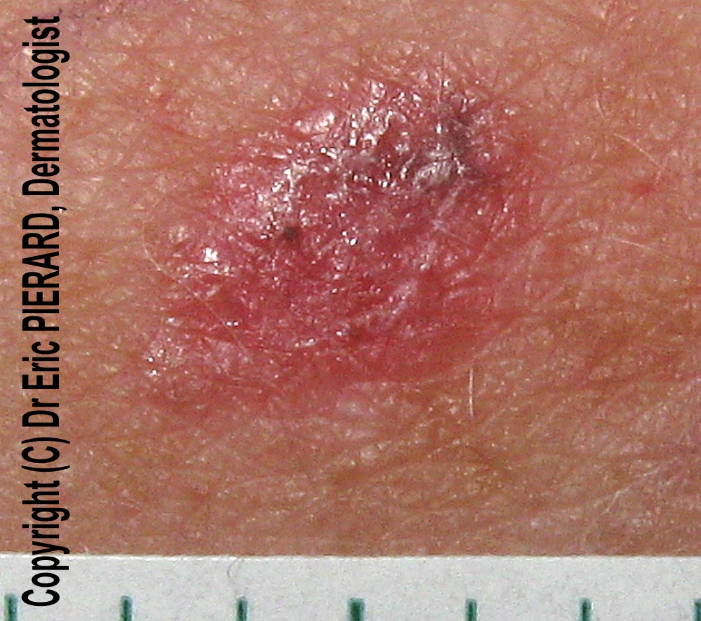
 A 50-year-old woman consulted for an enlarging lesion on her right arm;
Clinical examination revealed a red papular lesion with a slight pigmentation.


Dermoscopy revealed:

  •  atypical vascular pattern with polymorphous vessels, namely linear irregular vessels, dotted vessels
  • a small area of pigmentation (gray dots)
An excision was performed and pathology revealed an achromic melanoma with a Breslow index at 0.5mm.

Thursday, 26 March 2009

melanoma

A 82-year-old woman consulted for this dark pigmented lesion on her left arm. Clinically a melanoma was suspected.
Dermoscopy revealed:
  • a multi-coloured pattern
  • a blue white veil
  • blue gray dots
  • whitish scar-like depigmentation
  • polymorphous vessels including linear irregular vessels


Friday, 4 July 2008

Melanoma

Asymmetric lesion with pink homogeneous area

A 68-year old man consulted for an atypical pigmented lesion on his left lumbar area.

Dermoscopy revealed a melanocytic lesion characterized by:
  • asymmetry
  • an atypical pigment network
  • a pink homogeneous area
  • a white area of regression
  • linear-irregular vessels
  • polymorphous vessels
Polymorphous and linear-irregular vessels

Atypical pigment network (circle)

White area of regression (circle)

This lesion was highly suspicious of melanoma and excised.

Pathology revealed a superficial spreading melanoma (SSM) in situ developed on a melanocytic nevus developed on a melanocytic nevus

Saturday, 5 January 2008

Basal cell carcinoma

Clinical picture
A 44-year-old woman consulted for this lesion on her back with a circinar papulous border clinically suspicious of being a basal cell carcinoma.


Dermoscopy 1
In dermoscopy the lesion lacked pigment network.
There were blue- gray globules and polymorphous vessels (glomerular vessels, linear irregular vessels, hairpin vessels)

Dermoscopy 2


Dermoscopy 3
Glomerular vessel
and hairpin vessels (right part)


Dermoscopy 4
irregular linear vessels

In this case, vascular pattern was atypical for a basal cell carcinoma which features typically arborizing tree vessels. Irregular linear vessels are more often observed in melanomas.
The lesion was excised and pathology confirmed the diagnosis of basal cell carcinoma.


Other cases of basal cell carcinomas: 
1 2 3 4 5 6 7 8  10 11 12 13 14 15 16 17 18 19 20 21

Tuesday, 18 December 2007

Squamous cell carcinoma


A 70-year-old man consulted for a keratotic tumour on his scalp.


Dermoscopy revealed an unspecific pattern, with many polymorphous vessels.
Polymorphous vessels
were the only dermoscopic sign in favor of a malignant tumour.
Clinical and dermoscopic features were in favour of squamous cell carcinoma. A biopsy was performed and pathology confirmed this hypothesis.

Tuesday, 20 November 2007

Basal cell carcinoma

This 56-year-old man presented a basal cell carcinoma of his chest. The lesion had two parts: a red one at the left and a brown one at the right.
On the red part of the BCC, dermoscopy revealed arborizing vessels. On the brown part of the BCC, there were many polymorphous vessels and some blue-gray ovoid nests.