Search This Blog

Showing posts with label linear-irregular vessel. Show all posts

Wednesday, 4 June 2014

Regression


A 48-year-old woman presented this pigmented lesion on her back. 


 1: clinical picture

 2: dermoscopic picture

3: dermoscopic picture

  4:  dermoscopic picture


Dermoscopy revealed:
  • regression areas with some depigmentation (green arrows and ovoid area)
  • blue gray structures (blue arrows) and blue gray dots (peppering)
  • linear irregular vessels (black arrows)


Lesion was excised and pathology revealed a regressive superficial spreading melanoma with Breslow index at  0,39 mm

Friday, 23 March 2012

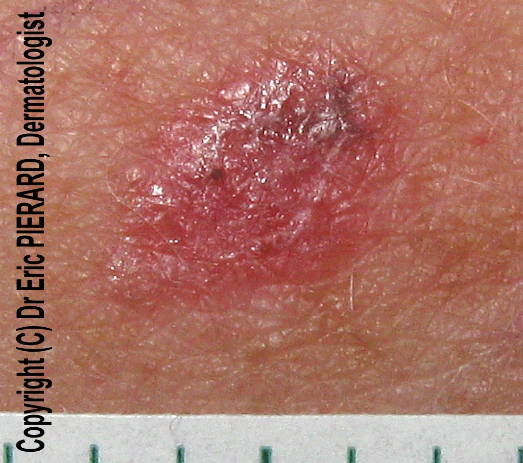
Red lesion on an arm...


 A 50-year-old woman consulted for an enlarging lesion on her right arm;
Clinical examination revealed a red papular lesion with a slight pigmentation.


Dermoscopy revealed:

  •  atypical vascular pattern with polymorphous vessels, namely linear irregular vessels, dotted vessels
  • a small area of pigmentation (gray dots)
An excision was performed and pathology revealed an achromic melanoma with a Breslow index at 0.5mm.

Thursday, 26 March 2009

melanoma

A 82-year-old woman consulted for this dark pigmented lesion on her left arm. Clinically a melanoma was suspected.
Dermoscopy revealed:
  • a multi-coloured pattern
  • a blue white veil
  • blue gray dots
  • whitish scar-like depigmentation
  • polymorphous vessels including linear irregular vessels


Friday, 4 July 2008

Melanoma

Asymmetric lesion with pink homogeneous area

A 68-year old man consulted for an atypical pigmented lesion on his left lumbar area.

Dermoscopy revealed a melanocytic lesion characterized by:
  • asymmetry
  • an atypical pigment network
  • a pink homogeneous area
  • a white area of regression
  • linear-irregular vessels
  • polymorphous vessels
Polymorphous and linear-irregular vessels

Atypical pigment network (circle)

White area of regression (circle)

This lesion was highly suspicious of melanoma and excised.

Pathology revealed a superficial spreading melanoma (SSM) in situ developed on a melanocytic nevus developed on a melanocytic nevus

Wednesday, 28 May 2008

Dermatofibroma with vascular structures


A 54-year-old woman consulted for a firm reddish tumor on her left thigh.
Dermoscopy revealed a central scarlike white patch in favor of a dermatofibroma and peripheral vascular structures: peripheral erythema and linear-irregular vessels.

peripheral erythema and linear-irregular vessels

Vascular structures are found in 29 to 49% of dermatofibromas, including erythema, dotted vessels, coma vessels, hairpin vessels, glomerular vessels, telangiectasias, linear-irregular vessels, polymorphous/atypical vessels.

Litterature:
Dermoscopy of dermatofibromas. A Prospective Morphological Study of 412 Cases
P. Zaballos, S. Puig, A. Llambrich, J. Malvehy, Arch Dermatol. 2008;144(1):75-83

Saturday, 10 May 2008

Atypical nevus


A 32-year-old woman consulted for a mole check-up.
Clinical examination revealed the greatest mole localized on her left scapular area.
At dermoscopy, this lesion featured a pink veil, irregular streaks, irregular globules, an atypical reticular pattern, linear irregular vessels.


Irregular globules.

Polymorphous vessels including irregular linear vessels.

Irregular streaks.

Two diagnoses were possible: superficial spreading melanoma or atypical nevus. The patient did not mention any changing in surface or borders.
This lesion was excised and pathology revealed an atypical nevus ( compound) with a slight irritation.

Saturday, 5 January 2008

Basal cell carcinoma

Clinical picture
A 44-year-old woman consulted for this lesion on her back with a circinar papulous border clinically suspicious of being a basal cell carcinoma.


Dermoscopy 1
In dermoscopy the lesion lacked pigment network.
There were blue- gray globules and polymorphous vessels (glomerular vessels, linear irregular vessels, hairpin vessels)

Dermoscopy 2


Dermoscopy 3
Glomerular vessel
and hairpin vessels (right part)


Dermoscopy 4
irregular linear vessels

In this case, vascular pattern was atypical for a basal cell carcinoma which features typically arborizing tree vessels. Irregular linear vessels are more often observed in melanomas.
The lesion was excised and pathology confirmed the diagnosis of basal cell carcinoma.


Other cases of basal cell carcinomas: 
1 2 3 4 5 6 7 8  10 11 12 13 14 15 16 17 18 19 20 21

Saturday, 27 October 2007

Squamous cell carcinoma

A 78-year-old man consulted for this rapidly enlarging tumor on the left side of his nose. The lesion was present for 3 months. The possible clinical diagnoses were keratoacanthoma, squamous cell carcinoma, and basal cell carcinoma.
Dermoscopy revealed whitish halo (A) and linear-irregular vessels (B). The lesion was excised and pathology was in favor of a squamous cell carcinoma.

Friday, 26 October 2007

Atypical dermatofibroma with vascular pattern

A 68-year-old man consulted for this lesion on his right leg present for more than 15 years.
Dermoscopy revealed some light brown globules (*) and a vascular pattern with polymorphous vessels: hairpin vessels, dotted vessels, and linear-irregular vessels.
A biopsy was performed and revealed a dermatofibroma.
Dermatofibromas with vascular pattern have been reported in the literature (D.Picolo, K.Peris, page 287, in color atlas of melanocytic lesions of the skin, Springer edition, 2007)