Search This Blog

Showing posts with label Basal cell carcinoma. Show all posts

Friday, 12 February 2016

A temporal lesion


A 60-year-old woman consulted for this temporal  pigmented lesion


 Dermoscopy revealed:

  • blue-gray dots / globules
  • leaf-like areas


Pathology was in favor of a basal cell carcinoma

Wednesday, 9 April 2014

Periocular blue nodule


A 28-year-old woman consulted for this slowly enlarging periocular tumor.


Dermoscopy revealed a bluish lesion with clear arborizing vessels.

This lesion was excised and pathology revealed a basal cell carcinoma.

Wednesday, 9 October 2013

Chrysalis structures


A 51-year-old patient consulted for this lesion located on his back.


Dermoscopy revealed :

  • arborizing vessels
  • ulcerations
  • chrysalis structures


This lesion was a basal cell carcinoma.


Crystalline or chrysalis structures are white shiny streaks that can only be seen with polarized dermatoscopes.

Crystaline or chrysalis structures are most commonly observed in basal cell carcinoma and invasive melanomas and rarely seen in nevi. In melanoma,they reflect increased tumor thickness and progression. 
The significance of crystalline/chrysalis structures in the diagnosis of melanocytic and nonmelanocytic lesions. Y. Balagula, R.P. Braun, H. S. Rabinovitz et al.  J Am Acad Dermatol 2012;67:194.e1-8.

Tuesday, 13 September 2011

A red tumor on a leg


A 42-year-old patient consulted for this red tumor present on his right leg for 1 year.


Dermoscopy revealed :

  • no pigment network
  • arborizing vessels
in favor of a basal cell carcinoma

Tuesday, 10 May 2011

Red nodule

A 62-year-old man consulted for a red tumor on his back.

Three dermoscopic pictures are exposed with different levels of pressure on the tumor. Vessels are well seen with a slight pressure.


Dermoscopy revealed arborizing vessels and a blue globule in favour of a basal cell carcinoma.

Tuesday, 1 February 2011

A facial telangiectatic tumor

A 67-year-old woman consulted for this small lesion on her left cheek.


Dermoscopy was typical of a basal cell carcinoma: ulceration, arborizing vessels and a (subtle) leaf -like area.

Wednesday, 18 August 2010

What is this lesion on a leg (first case) ?


A 72-year-old woman consulted for this ulcerated lesion on her right leg.

Dermoscopy revealed:
  • ulceration (crusts)
  • central glomerular vessels regularly disposed
  • blue-gray globules
  • spokewheel areas
Discussion: blue-gray globules were the main sign in favor of a basal cell carcinoma (BCC), in association with ulceration and some spokewheel areas.
Nevertheless, glomerular vessels have not been reported in BCC. We think that, in our case, these glomerular vessels were due to a stasis dermatitis.

An excision was performed and pathology confirmed the diagnosis of BCC.

Monday, 16 August 2010

Nasal lesion.

A 72-year-old man consulted for a lesion on his nose.

Dermoscopy revealed:
  • absence of pigment network
  • arborizing blood vessels
  • blue-gray globules
  • spokewheel areas
in favor of a basal cell carcinoma and confirming the clinical hypothesis.

Wednesday, 28 April 2010

Basal cell carcinoma


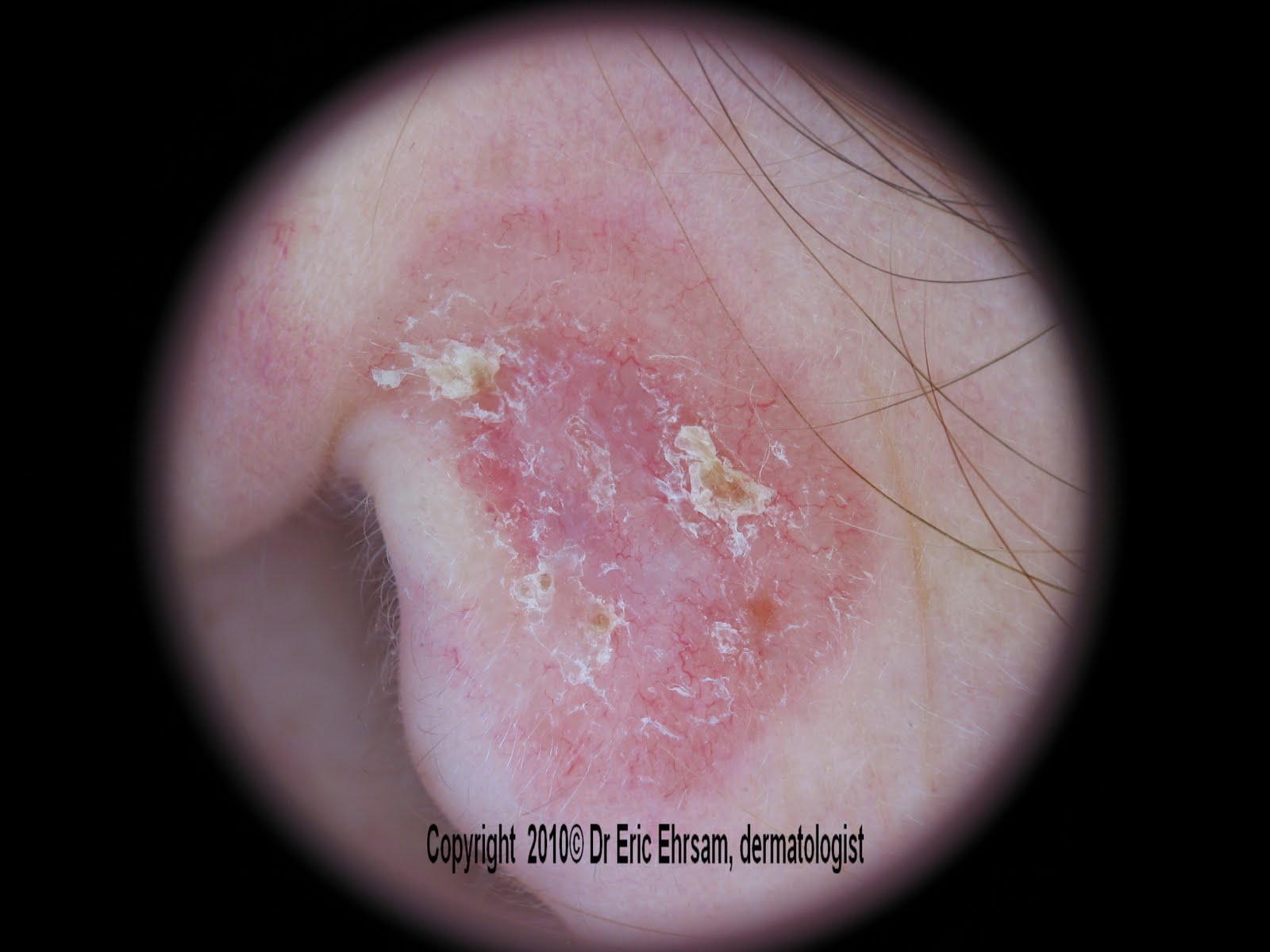
35-year-old woman with an enlarging scaly lesion.


Arborizing vessels were typically in favor of a basal cell carcinoma.


Thursday, 11 June 2009

Leaves


A 48-year-old woman presented a pigmented lesion on her sternal area:


Dermoscopy revealed:
  • a non melanocytic pattern
  • typical leaf-like structures
  • one spoke wheel-like structure
in favour of a basal cell carcinoma


Typical leaf-like areas (click on the picture above to see enlargement)

Sunday, 10 May 2009

A scalp tumour...

A 38-year-old woman presented since birth a localised parietal hairless orange-yellow plaque (1) This plaque was smooth and became verrucous during childhood.
The patient consulted because of the occurence of nodules (2) on the anterior part of the lesion.

Dermoscopy revealed yellow globular-like structures (1) on the posterior area.
On the nodules, arborizing tree vessels and blue ovoid nests were in favour of a basal cell carcinoma or a trichoblastoma
This lesion was biopsied  and pathology confirmed the diagnosis of basal cell carcinoma occuring on a Jadahsson nevus (nevus sebaceus)
Knowing that trichoblastomas are the tumors the most frequently reported as complication of nevus sebaceous  (2, 3) and that dermoscopy is not sufficently discriminative to distinguish a trichoblastoma from a basal cell carcinoma, we have to wait for the pathology result of the entire excision.
An update will be added within a month.

Litterature

1 -Nancy H. Kim et al.  The dermoscopic differential diagnosis of yellow lobularlike structures. Arch Dermatol 2008; 144 (7): 962.

2-Jaqueti et Al. Trichobastoma is the most common neoplasm debeloped in nevus sebaceus of Jadassohn: a clinicopathologic study of a series of 155 cases. Am J Dermatopathol 2000;22(2):108-18

3- Cribier et Al. Tumors arising in nevus sebaceus. A study of 596 cases. J Am Acad Dermatol 2000;42:263-8


Wednesday, 10 December 2008

Blue-gray globules

A 67-year-old woman consulted for this lesion on her back. Dermoscopy revealed blue-gray globules (rectangle) and some arborizing vessels (arrows) confirming easily the diagnosis of basal cell carcinoma.


Blue-gray globules are a pathognomonic sign  in pigmented basal cell carcinoma. 
They are often associate with other basal cell carcinoma specific criteria, as leaf-like structures,
spoke-wheel areas and  large blue-gray ovoid nests.

Saturday, 6 December 2008

Arborizing tree vessels

A 54-year-old woman consulted for this small pink lesion on her scalp (parietal area)


Dermoscopy revealed many arborizing vessels in favour of a basal cell carcinoma.

The lesion was excised and pathology confirmed the clinical and dermoscopic diagnoses.


Monday, 8 September 2008

Leaf-like structures

A 92-year-old woman presented a red tumor on her neck.


Dermoscopy revealed many brown structures with a leaf-like configuration located at the periphery of the lesion. This was typical of leaf-like structures, also called maple leaf-like areas in the first publications.

Leaf-like structures are quite exclusively found in basal cell carcinomas but have also been reported in featureless melanomas.

Other cases of basal cell carcinomas: 
1 2 3 4 5 6 7 8  10 11 12 13 14 15 16 17 18 19 20 21

Friday, 29 August 2008

Basal cell carcinoma


A 72-year-old woman consulted for this pigmented lesion on the left side of her nose.

Dermoscopy revealed a non melanocytic tumor with:
  • large blue ovoïd nest
  • arborizing vessels (in the blue ovoïd nest)
  • blue gray globules
  • leaf-like areas
in favor of a basal cell carcinoma.


leaf-like structures

Other cases of basal cell carcinomas: 
1 2 3 4 5 6 7 8  10 11 12 13 14 15 16 17 18 19 20 21

Sunday, 27 July 2008

Basal cell carcinoma

A 72-year-old woman consulted for this red left cervical tumor.


Dermoscopy revealed an unspecific pattern and many arborizing blood vessels in favor of a basal cell carcinoma.

Arborizing vessels are the most typical local criteria in basal cell carcinoma.


They are rarely observed in other tumors:
  • melanocytic nevus
  • melanoma
  • seborrheic keratosis
Other cases of basal cell carcinomas: 
1 2 3 4 5 6 7 8  10 11 12 13 14 15 16 17 18 19 20 21

Tuesday, 24 June 2008

Basal cell carcinoma

A 59-year-old man consulted for this tumor on his nose.

Dermoscopy was typically in favor of a basal cell carcinoma:
  • arborizing blood vessels
  • blue ovoid nest
  • blue gray globules
  • ulceration with congealed blood
Other cases of basal cell carcinomas: 
1 2 3 4 5 6 7 8  10 11 12 13 14 15 16 17 18 19 20 21

Saturday, 21 June 2008

Basal cell carcinoma

A 89-year-old woman consulted for a pigmented lesion on her forehead. A basal cell carcinoma was the main clinical diagnosis.

On this picture, with a slight pressure of the dermoscope (polarized light dermoscopy), arborizing blood vessels are well seen.
Blue gray dots and globules are the other dermoscopic signs in favor of this basal cell carcinoma.

If the pressure of the dermoscope is too important, the blood vessels are not visible.

Vessels and red areas are better visualized with polarized light dermoscopy than with immersion contact dermoscopy (1)

References:
1 - Marghoob et al. Differences between polarized light dermoscopy and immersion contact dermoscopy for the evaluation of skin lesions. Arch Dermatol 2007 Mar;143(3):329-38