Search This Blog

Showing posts with label globule. Show all posts

Tuesday, 24 August 2010

Pigmented breast lesion

A 28-year-old woman consulted for an enlarging pigmented lesion on her left breast.

Dermoscopy revealed:
  • a multi-colored pattern
  • a multicomponent pattern
  • asymmetry in 2 axes
  • an atypical reticular pattern
  • some atypical dots/globules
  • a scar-like depigmentation
  • a blue white veil
Pathology revealed a superficial spreading melanoma with a Breslow index at 0,5mm

Saturday, 1 August 2009

Spitz nevus


A 23-year-old woman consulted for this pigmented lesion in her back.
Its history was unclear.
Clinically this lesion was an ugly duckling by comparison with other moles she had.

Dermoscopy revealed a reticular pattern, irregular globules especially visible at the periphery and some pseudopods.

Dermoscopic presentation was in favor of a Spitz nevus.
The lesion was excised and pathology confirmed the diagnosis.


Wednesday, 9 July 2008

Halo nevus

A 16-year-old boy underwent a yearly mole check-up. One year later, one of his nevi presented a peripheral depigmentation in favor of halo nevus.


The difference between the two dermoscopic images at a one year interval was very marked:
  • depigmentation of the surrounding skin
  • enlightening of the global pattern and rarefaction of the globules

In this case occuring in a teen, there were no atypical dermoscopic features in favor of a melanoma and another appointment was scheduled one year later.


Halo nevus is a benign lesion and no treatment is required. It is a frequent phenomena during childhood. Despite benign features, the presence of a new halo lesion in an adult has to be examined with a high index of suspcion of melanoma. If a halo nevus is showing atypical dermoscopic features, an excision has to be scheduled.

Wednesday, 2 January 2008

Compound melanocytic nevus


A 14-year-old girl consulted for an enlarging melanocytic nevus on her back. One of her sister, aged 24, had been operated for a superficial spreading melanoma 2 years before, and she was very anxious. The lesion was larger than 6 mm and had an irregular border.


Dermoscopy revealed a central homogeneous pattern and a peripheral typical reticular pattern, and peripheral globules, but no signs for a SSM.
The lesion was removed and pathology was in favour of a compound melanocytic nevus.

Friday, 21 December 2007

Halo nevus

A 25-year-old man consulted for 2 cases of depigmentation around nevi on his back.


Case 1: the nevus became lighter and there was a rim of depigmentation


Case 1: dermoscopy revealed a globular pattern with globules barely visible

Case 2: the globules disappeared and there was only a depigmentation


Keywords: halo nevus, Sutton nevus, leukoderma acquisitum centrifugum, depigmentation, globular pattern.

Thursday, 13 December 2007

Congenital nevus

A 12-year-boy consulted for the follow-up of a congenital melanocytic nevus on his abdomen.

Dermoscopy revealed a typical reticular pattern and regular globules.

Monday, 3 December 2007

Rim of globules


A 16-year-old boy consulted for his nevi after summer holidays. Dermoscopy revealed 3 nevi with rim of globules. This dermoscopic feature is often seen after sun exposition and in enlarging nevi.


Spitz nevus


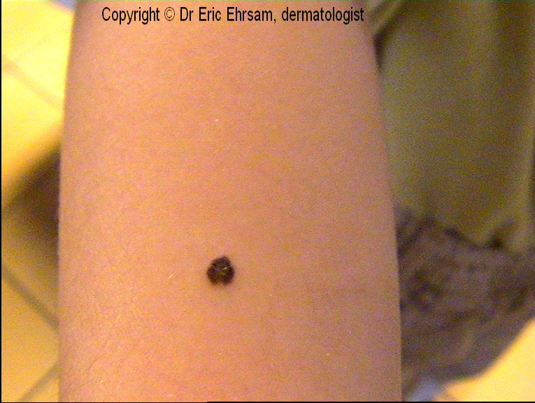
11-year-old boy with a dark pigmented lesion on his right arm.
Dermoscopy: symetric starburst pattern, streaks, regular globules at the center of the lesion. The diagnosis of Spitz nevus was made and a dermoscopic follow-up did not reveal enlargement or changing of structures.


Thursday, 29 November 2007

Dermatofibroma

A 35-year-old woman consulted for a painful firm pigmented tumor clinically in favor of a dermatofibroma.


Dermoscopy revealed a central white scar-like patch surrounded by a pigmented network and many brown globules.


Dermatofibromas have various dermoscopic features.

For more information:

Isil Kilinc Karaarslan et al. Different dermoscopic faces of dermatofibromas. J Am Acad Dermatol 2007;57:401-6

Other cases of dermatofibromas
1 2 3 4 5 6 7 8 9 10 11 12


Tuesday, 27 November 2007

Superficial spreading melanoma


A 60-year-old man consulted for a changing of color of one of his nevi on his back. The upper part of this nevus turned dark brown a few months before.


On the upper part of the lesion, dermoscopy revealed an atypical reticular pattern, asymmetric irregular globules and a blotch.


On the lower part of the lesion, there was an homogeneous non pigmented pattern with some comma vessels in favor of a dermal nevus.


The tumor was excised and pathology revealed a superficial spreading melanoma developed on a pre-existing dermal nevus.

Other cases of melanoma
0 1 2 3 4 5 6 7 8 9 10 11 12 13 14 15 16 17 18

Saturday, 24 November 2007

Spitz nevus

A 15-year-old boy presented a Spitz nevus confirmed by excision.
The lesion was frankly pigmented and there was a rim of brown to black globules at the periphery.
This dermoscopic aspect was different from classic cases of pigmented Spitz nevi where the peripheral globules are fused with the central body of the nevus realising streaks in a starburst pattern.


Sunday, 18 November 2007

Eccentric hyperpigmented area

This pigmented lesion was asymetrically pigmented. Dermoscopy revealed a globular pattern in the center and a reticular pattern at the periphery with an eccentric hyperpigmented area (circle)
There were no dermoscopic criteria for a melanoma. We have to be very careful with this dermoscopic feature of eccentric hyperpigmented area because it is observed in atypical nevus but also in melanoma in situ and early invasive melanoma. Two attitudes could have been proposed: 3-month dermoscopic follow-up or immediate excision. In our case we choose to cut it off and pathology revealed a benign compound nevus (dermal and junctional

Saturday, 17 November 2007

Spitz nevus

A 14-year-old boy presented a dark pigmented lesion on his lumbar area. Dermoscopy revealed a homogeneous pattern with a bluish pigmentation. Rare brown globules were found at the periphery. The lesion was excised and pathology revealed a dermal Spitz nevus.

Six dermoscopic patterns of Spitz nevus have been described:
-vascular pattern with dotted vessels
-globular pattern
-reticular pattern
-starburst pattern
-homogeneous pattern
-atypical pattern

Friday, 26 October 2007

Atypical dermatofibroma with vascular pattern

A 68-year-old man consulted for this lesion on his right leg present for more than 15 years.
Dermoscopy revealed some light brown globules (*) and a vascular pattern with polymorphous vessels: hairpin vessels, dotted vessels, and linear-irregular vessels.
A biopsy was performed and revealed a dermatofibroma.
Dermatofibromas with vascular pattern have been reported in the literature (D.Picolo, K.Peris, page 287, in color atlas of melanocytic lesions of the skin, Springer edition, 2007)


Thursday, 18 October 2007

Congenital melanocytic nevus

This 17-month-old boy presented a typical congenital melanocytic nevus with an homogenous globular pattern.

Saturday, 11 August 2007

superficial spreading melanoma


A 84-year-old man presenting many seborrheic keratoses, developed a slowly enlarging pigmented lesion of the abbdomen


On dermoscopy (x20): asymetric lesion with atypical globular pattern, irregular dots and globules at 3 o clock (right side) , and blue white veil


The lesion was removed and pathologist concluded to a superficial spreading melanoma Breslow index 0.28mm


Other cases of melanoma
0 1 2 3 4 5 6 7 8 9 10 11 12 13 14 15 16 17 18