Search This Blog

Showing posts with label melanocytic nevus (Spitz). Show all posts

Saturday, 28 November 2015

Black lesion.


A 25-year-old woman consulted for a dark pigmented lesion on her abdomen


Dermoscopy revealed:

  • asymmetry of form
  • a central blotch
  • regular pseudopods
Pathology was in favor of melanocytic Spitz nevus

Sunday, 30 March 2014

Acquired lesion on an ankle.

A 26-year-old man consulted for an acquired lesion for 4 years which was slowly enlarging on his right ankle.


 Clinical picture.


 Dermoscopic picture without pressure.


Dermoscopic picture with pressure.

This regular lesion has:
  • regular dotted vessels
  • a retiform depigmentation, or reticular depigmentation  
Clinical and dermoscopic patterns were in favor of a Spitz nevus.
Pathology confirmed this diagnosis.


Tuesday, 13 September 2011

A red tumor on a leg


A 42-year-old patient consulted for this red tumor present on his right leg for 1 year.


Dermoscopy revealed :

  • no pigment network
  • arborizing vessels
in favor of a basal cell carcinoma

Monday, 4 July 2011


A 25-year-old man consulted presented a pigmented lesion on his back (lumbar area).


Dermoscopy revealed:

  • a réticular pattern
  • irregular streaks (pseudopods)
in favor of a Spitz nevus which was confirmed after excision.

Saturday, 1 August 2009

Spitz nevus


A 23-year-old woman consulted for this pigmented lesion in her back.
Its history was unclear.
Clinically this lesion was an ugly duckling by comparison with other moles she had.

Dermoscopy revealed a reticular pattern, irregular globules especially visible at the periphery and some pseudopods.

Dermoscopic presentation was in favor of a Spitz nevus.
The lesion was excised and pathology confirmed the diagnosis.


Monday, 3 December 2007

Spitz nevus


11-year-old boy with a dark pigmented lesion on his right arm.
Dermoscopy: symetric starburst pattern, streaks, regular globules at the center of the lesion. The diagnosis of Spitz nevus was made and a dermoscopic follow-up did not reveal enlargement or changing of structures.


Saturday, 24 November 2007

Spitz nevus

A 15-year-old boy presented a Spitz nevus confirmed by excision.
The lesion was frankly pigmented and there was a rim of brown to black globules at the periphery.
This dermoscopic aspect was different from classic cases of pigmented Spitz nevi where the peripheral globules are fused with the central body of the nevus realising streaks in a starburst pattern.


Monday, 19 November 2007

Spitz nevus

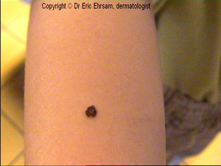
A 15-year-girl consulted for a dark pigmented lesion on her left arm. Dermoscopy revealed a typical symetric starburst pattern in favor of a Spitz/Reed nevus
The lesion was excised and pathology confirmed the diagnosis.

Sunday, 18 November 2007

Spitz nevus

5-year-old boy with pink tumor on his right cheek clinically in favor of a Spitz nevus.
Dermoscopy revealed a non specific regular pseudo-network with a granular appearance.
Dermoscopy criteria are useful for pigmented Spitz nevi or Reed nevi.
In this case, a regular 6-months follow-up was proposed.

Saturday, 17 November 2007

Spitz nevus

A 14-year-old boy presented a dark pigmented lesion on his lumbar area. Dermoscopy revealed a homogeneous pattern with a bluish pigmentation. Rare brown globules were found at the periphery. The lesion was excised and pathology revealed a dermal Spitz nevus.

Six dermoscopic patterns of Spitz nevus have been described:
-vascular pattern with dotted vessels
-globular pattern
-reticular pattern
-starburst pattern
-homogeneous pattern
-atypical pattern

Wednesday, 7 November 2007

Spitz nevus

A 40-year-old woman consulted for this slowly enlarging lesion on her right buttock. Dermoscopy revealed a starburst pattern and a slight blue white veil.
All spitzoïd lesion of adulthood has to be excised.
In this case dermoscopy revealed a pigmented Spitz nevus

Thursday, 18 October 2007

Spitz nevus

A 32-year-old woman consulted for this pigmented lesion on her left wrist. The lesion was excised. Pathology revealed a combined Spitz nevus.


Dermoscopy showed a negative pigment network also called retiform depigmentation

Wednesday, 3 October 2007

Starburst pattern

A 10-year-old girl consulted for this black lesion on her back.
Dermoscopy revealed a typical regular starburst pattern with pseudopods regularly distributed at the periphery in favor of a benign Reed nevus.
A regular follow-up was planned.


Tuesday, 28 August 2007

Spitz nevus


A 47-year-old woman consulted for a a slowly enlarging pigmented lesion of her right wrist for 3 months.


Dermoscopy (x20) revealed regular streaks throughout the lesion (pseudopods and radial streaming) with a symmetrical starburst pattern.

Because of the age of the patient and the rapid enlargement, the lesion was removed.

Pathology revealed a Spitz nevus