Search This Blog

Showing posts with label dotted vessels. Show all posts

Sunday, 30 March 2014

Acquired lesion on an ankle.

A 26-year-old man consulted for an acquired lesion for 4 years which was slowly enlarging on his right ankle.


 Clinical picture.


 Dermoscopic picture without pressure.


Dermoscopic picture with pressure.

This regular lesion has:
  • regular dotted vessels
  • a retiform depigmentation, or reticular depigmentation  
Clinical and dermoscopic patterns were in favor of a Spitz nevus.
Pathology confirmed this diagnosis.


Friday, 23 March 2012

Red lesion on an arm...


 A 50-year-old woman consulted for an enlarging lesion on her right arm;
Clinical examination revealed a red papular lesion with a slight pigmentation.


Dermoscopy revealed:

  •  atypical vascular pattern with polymorphous vessels, namely linear irregular vessels, dotted vessels
  • a small area of pigmentation (gray dots)
An excision was performed and pathology revealed an achromic melanoma with a Breslow index at 0.5mm.

Tuesday, 13 September 2011

A red tumor on a thigh


A 3-year-girl was brought for examination of this non pigmented lesioon on her thigh.


Dermoscopy (X20) revealed a vascular pattern with predominance of dotted vessels.


Same image with a heavier pressure on dermoscope: vessels are less visible.

Clinical and dermoscopic images were in favor of a Spitz nevus.

Thursday, 15 January 2009

Clear cell hidradenoma

Fig 1: clinical picture

A 52-year-old man consulted for a scapular nodule. Clinical examination revealed a flesh-coloured nodule (Fig 1)

Fig 2 dotted vessels

Polarizing dermoscopy with a slight pressure revealed dotted vessels (Fig 2)

Fig 3 whitish areas

Fig 4 whitish areas

Dermoscopy with a louder pressure revealed whitish areas (Fig 3 and 4).

This lesion was excised and pathology revealed a clear cell hidradenoma.

Clear cell hidradenomas are usually benign intradermal tumors and represent 95% of all hidradenomas. These tumors have a slow growth and sometimes can be ulcerated. Theay have to be surgically excised because of a high risk of recurrence and a potential of malignant evolution.

Y. Yoshida and al. reported dermoscopic features of a clear cell hidradenoma (Y. Yoshida and al. Dermatology 2008;217: 250-251).
They found in their case whitish areas, reddish purple areas, and some linear or hairpin vessels on the surface of the tumour. Reddish purple areas were corresponding to cystic spaces with hemorrhage.

Friday, 20 June 2008

Atypical dermatofibroma

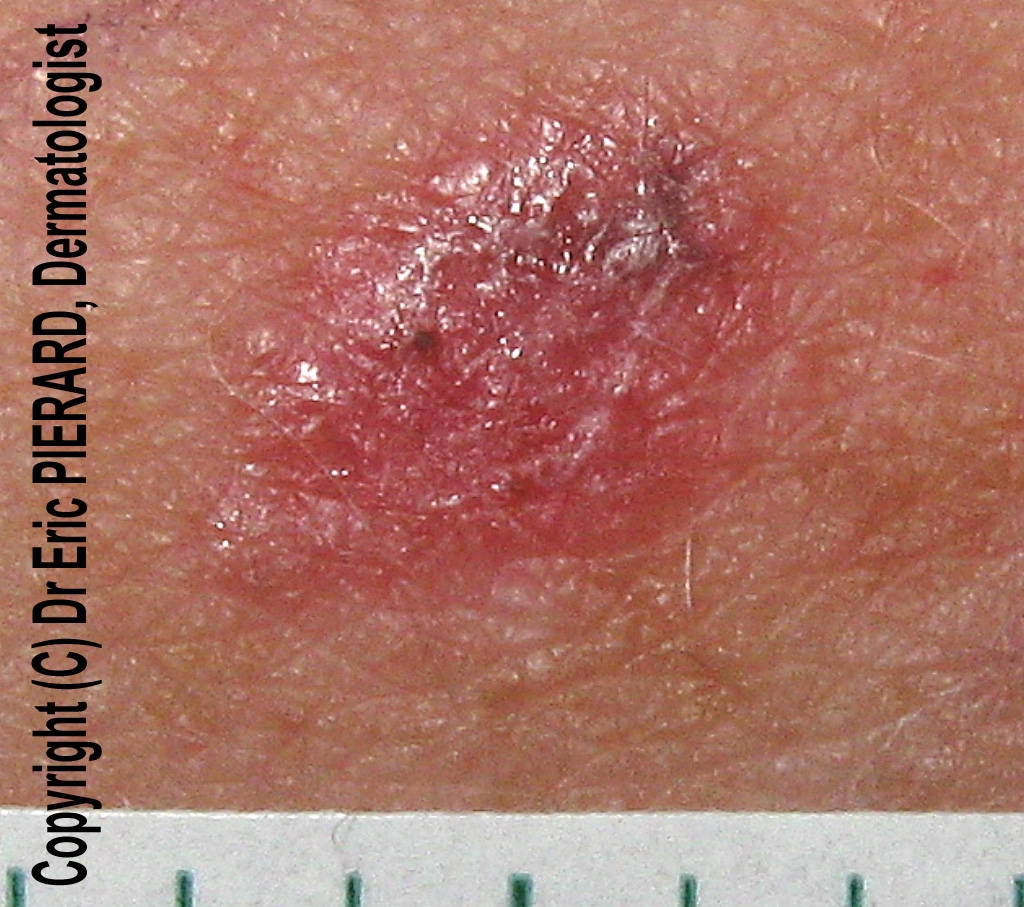
A 27-year-old woman consulted for a red firm lesion on her right cheek. This lesion was present for 1 year.

Picture 1: slight pressure of the dermoscope

Dermoscopy revealed a dotted vascular pattern (dotted vessels) as the only dermoscopic sign.
Clinical diagnoses were Spitz nevus, hypopigmented Clark nevus and amelanotic melanoma. Dotted vessels are typically reported in these 3 cutaneous tumors.

The lesion was excised and pathology revealed a dermatofibroma.

Picture 2: more pressure of the dermoscope

Similar cases of dermatofibroma with a dotted vascular pattern are rarely reported in the literature (1- 2 - 3)

Dermatofibromas are rare on the face. Cases of of dermatofibromas of the face have to be excised with wider margins in comparison with examples of classical dermatofibromas occuring on the extremities because of diffuse infiltration, involvement of deeper structures and a increased rate of local recurrences (4)


References:

1 - Color atlas of melanocytic lesions of the skin. Soyer et al. Ed Springer. Chapter V.2: Piccolo and Peris: page 287

2 - Pedro Zaballos et al. Dermoscopy of Dermatofibromas. A Prospective Morphological Study of 412 CasesArch Dermatol. 2008;144(1):75-83

3 - Ferrari A et al. Cutaneous amelanotic melanoma metastasis and dermatofibromas showing a dotted vascular pattern. Acta Derm Venereol. 2004;84(2):164-165.

4 - Mentzel et al. Benign fibrous histicocytoma (dermatofibroma) of the face. Clinicopathologic and immunohistochemical study of 34 cases associated with an aggressive clinical course. Am J Dermatopathol 2001; 23(5): 419 -26

Wednesday, 11 June 2008

Pink nevus


A 25-year-old woman with a phototype I consulted for a check-up of many nevi on her trunk and limbs. Two nevi were pink at the difference of the majority of them which were light brown.
Dermoscopy revealed dotted vessels disposed in a regular pattern.
Pink nevi
are typicallly found in patients with fair skin type (phototypes I and II). If there are multiple pink lesions, a regular follow-up is advised as it is in this case.
If the lesion is solitary, it is better advised to remove it because differentiation from an achromic melanoma is not possible without pathology examination.


Other cases of pink nevi: 1

Litterature: Zalaudek and al, in Color atlas of melanocytic lesions of the skin. Springer Ed, Berlin, 2007


Friday, 1 February 2008

Wart


Dermoscopy may be sometimes useful to confirm the diagnosis of wart, when it is clinically atypical. In this case, the verruca vulgaris, located on the foot, was pink.
Dermoscopy showed dotted vessels surrounded by a whitish halo


Friday, 23 November 2007

Pink nevus

A 31-year-old woman with a phototype II consulted for this pink tumor on her right thigh. The lesion had enlarged for a couple of months but the patient did not remember exactly when it had occured. Clinical examination was in favor of a Spitz nevus, differential diagnoses included achromic melanoma, clear cell acanthoma, dysplastic nevus.
Dermoscopy revealed dotted vessels and a light brown color at the the periphery. The lesion was excised and pathology revealed a compound melanocytic nevus.
Pink nevi
are typicallly found in patients with fair skin type (phototypes I and II). If there are multiple pink lesions, a regular follow-up is advised. If the lesion is solitary as in our case, differentiation from an achromic melanoma is not possible and the lesion has to but excised for pathology.

Ref. Zalaudek and al, in Color atlas of melanocytic lesions of the skin. Springer Ed, Berlin, 2007


Friday, 26 October 2007

Atypical dermatofibroma with vascular pattern

A 68-year-old man consulted for this lesion on his right leg present for more than 15 years.
Dermoscopy revealed some light brown globules (*) and a vascular pattern with polymorphous vessels: hairpin vessels, dotted vessels, and linear-irregular vessels.
A biopsy was performed and revealed a dermatofibroma.
Dermatofibromas with vascular pattern have been reported in the literature (D.Picolo, K.Peris, page 287, in color atlas of melanocytic lesions of the skin, Springer edition, 2007)