Search This Blog

Showing posts with label blotch. Show all posts

Friday, 12 February 2016

Brachial melanoma


A 54-year-old man had a rapidly growing lesion on his right back for 6 months.


Dermoscopy revealed:
  • a vascular pattern with some polymorphous and atypical vessels
  • peripheral pigmentation
  • irregular globules
  • eccentric blotches


Pathology revealed a superficial spreading melanoma with Breslow index at 1.53 mm

Saturday, 16 January 2016

Small, beware !


A 42-year-old man consulted for this small (4mm) pigmented lesion on his left calf.


Dermoscoy revealed:

  • asymmetry of colors and structures
  • some irregular blotches
  • an atypical pigment network
  • a radial streaming


Lesion was excised and pathology was in favor or a melanoma in situ.

Saturday, 28 November 2015

Black lesion.


A 25-year-old woman consulted for a dark pigmented lesion on her abdomen


Dermoscopy revealed:

  • asymmetry of form
  • a central blotch
  • regular pseudopods
Pathology was in favor of melanocytic Spitz nevus

Saturday, 8 November 2014

Starburst pattern


A 43-year-old man consulted for this acquired pigmented lesion on his left leg.


Dermoscopy revealed:
  • a central blotch
  • a regular starburst pattern


 Two diagnoses were evoked: Reed nevus or melanoma. Regular blotch and symmetrical starburst pattern favored Reed nevus.

Lesion was excised and pathology revealed a superficial spreading melanoma with a Breslow index at 0.28mm.

Sunday, 7 September 2014


A 32-year-old man consulted for this enlarging pigmented lesion.


Clinical presentation: multiple couleurs, asymmetry

Dermoscopy revealed:

  • multiple colors
  • irregular dots
  • pink structureless area
  • irregular blotch
  • regression
This lesion was easily diagnosed superficial spreading melanoma.

Pathology revealed a superficial spreading melanoma with a Breslow index at 0.5mm

Tuesday, 23 August 2011

Pectoral lesion


A 52-year-old man presented this pectoral lesion. No clinical history was available.


Dermoscopy revealed:

  • multiple colours
  • a multi-component pattern
  • an atypical reticular pattern
  • some pseudopods 
  • irregular blotches
  • regression areas
Pathology confirmed the diagnosis of superficial spreading melanoma.

Friday, 22 July 2011

Back lesion


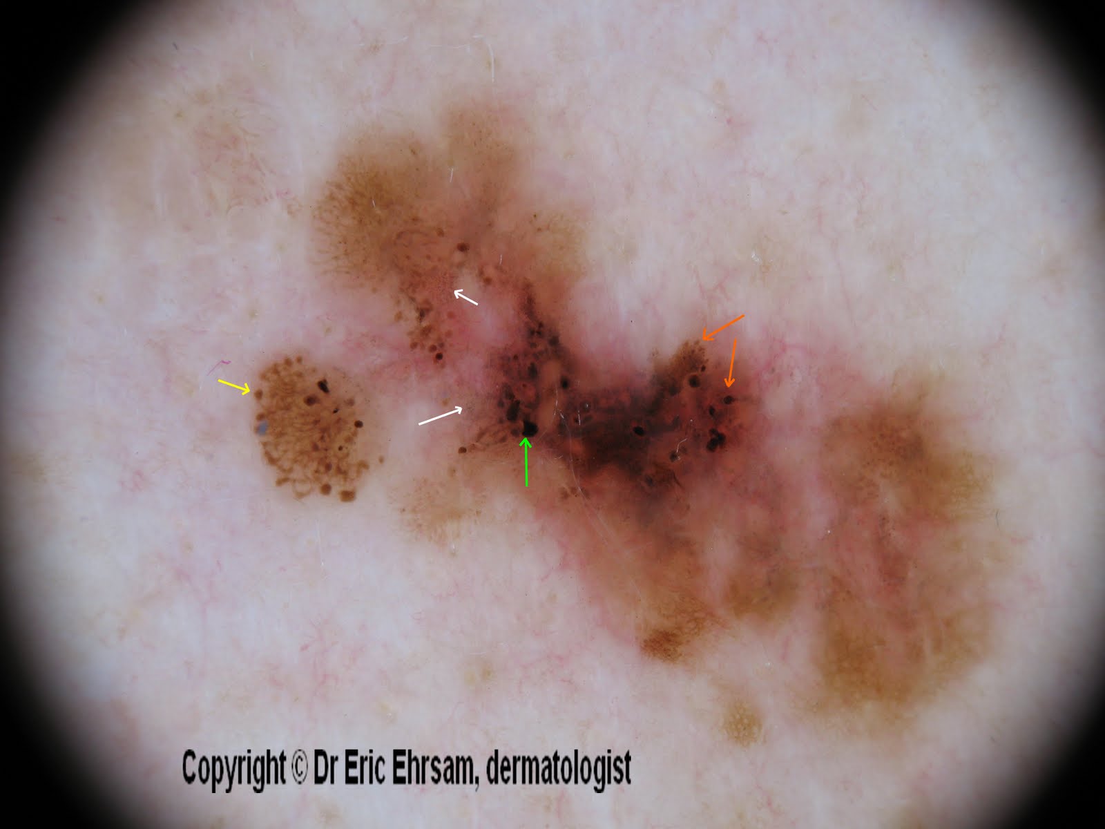
A 54-year-old woman consulted for this changing lesion on her back.


Dermoscopy revealed:

  • multiple colours, irregular pigmentation
  • a multi-component pattern
  • an atypical reticular pattern
  • irregular streaks (yellow arrow)
  • irregular dots and globules (orange arrow)
  • irregular blotches (green arrow)
  • peppering, regression (white arrow)


Pathology revealed a superficial spreading melanoma (SSM) with a Breslow index at 0.33 mm

Tuesday, 5 April 2011

Scalp lesion

A 72-year-old man consulted for a rapidly growing black nodule on his scalp. (the scar is due to an operation 3 years before for a Bowen's disease).

Dermoscopy revealed a dark blotch. At the border, a blue white veil is present.

This lesion was excised and pathology revealed a nodular melanoma (Breslow index 2 mm)

Friday, 21 January 2011

Black tumor


A 79-year-old man with many dysplastic nevi, consulted for a nodular bleeding tumour on his back.

Dermoscopic examination was difficult because this lesion was bleeding.
Dermoscopy revealed a bluish pattern, blotches and a brown pigmentation at the border of this lesion.
Pathology revealed a nodular melanoma (Breslow index 3,3 mm).

Wednesday, 18 August 2010

What is this lesion on a leg (2nd case) ?


A 71-year old woman consulted for this enlarging multi-colored lesion on her right leg.

Dermoscopy revealed:
  • irregular pigmentation
  • atypical pigment network
  • negative pigment network
  • irregularly distributed blotch
  • blue-white veil
in favor of a melanoma.

An excision was performed and pathology revealed a superficial spreading melanoma in vertical extension, Clark level IV, and Breslow index at 3,1 mm.

Sunday, 18 January 2009

Symmetric blotch

Fig 1 Blotch

A blotch (fig 1)  is a dark-brown or black structure-less area where other dermoscopic features cannot be seen. Symmetric blotches are in favour of benign lesions. Assymetrically distributed blotches are in favour of melanomas.
In our case, the blotch was symmetrically disposed and a peripheral reticular network was visible. This kind of blotch is also called black lamella.
This lesion was corresponding to a benign melanocytic nevus.

It has to be distinguished from the blue structure-less pattern or blue homogeneous pattern of a blue nevus (fig 2).

Fig 2 Blue homogeneous pattern

Saturday, 3 May 2008

Blotch


Blotch (or black lamella) is a black or dark brown pigmentation.
Blotch is formed by a large quantity of melanin pigment in the epidermis and / or dermis.
Here is a case of melanocytic junctional nevus where a blotch is covering its largest area.
In malanomas, blotches are often irregular.

Tuesday, 27 November 2007

Superficial spreading melanoma


A 60-year-old man consulted for a changing of color of one of his nevi on his back. The upper part of this nevus turned dark brown a few months before.


On the upper part of the lesion, dermoscopy revealed an atypical reticular pattern, asymmetric irregular globules and a blotch.


On the lower part of the lesion, there was an homogeneous non pigmented pattern with some comma vessels in favor of a dermal nevus.


The tumor was excised and pathology revealed a superficial spreading melanoma developed on a pre-existing dermal nevus.

Other cases of melanoma
0 1 2 3 4 5 6 7 8 9 10 11 12 13 14 15 16 17 18