Search This Blog

Friday 21 December 2007

Halo nevus

A 25-year-old man consulted for 2 cases of depigmentation around nevi on his back.


Case 1: the nevus became lighter and there was a rim of depigmentation


Case 1: dermoscopy revealed a globular pattern with globules barely visible

Case 2: the globules disappeared and there was only a depigmentation


Keywords: halo nevus, Sutton nevus, leukoderma acquisitum centrifugum, depigmentation, globular pattern.

Wednesday 19 December 2007

Nevus spilus

A 25-year-old woman consulted for a pigmented lesion on her right arm.
The lesion was a lentiginous patch with darker brown pigmented macules in favor of a nevus spilus.
Dermoscopy revealed darker brown areas with a reticulo-globular pattern in favor of melanocytic nevi. The background was light brown and reticular.

Nevus spilus are present at birth or childhood. On histology they correspond to a lentigo with surimposed junctional or compound nevi.
Main differential diagnosis is agminated nevus which correspond to lentiginous compound melanocytic nevi without light background pigmentation.

Reference: Bragg et al. Agminated acquired melanocytic nevi of the common and dysplastic type. J Am Acad Dermatol 2005;52:67-73


Tuesday 18 December 2007

Squamous cell carcinoma


A 70-year-old man consulted for a keratotic tumour on his scalp.


Dermoscopy revealed an unspecific pattern, with many polymorphous vessels.
Polymorphous vessels
were the only dermoscopic sign in favor of a malignant tumour.
Clinical and dermoscopic features were in favour of squamous cell carcinoma. A biopsy was performed and pathology confirmed this hypothesis.

Sunday 16 December 2007

Parallel ridge pattern


A 27-year-old woman consulted for this pigmented lesion on the sole of her left foot present for 2 months. Dermoscopy revealed a parallel ridge pattern and the presence of aggregated black globules, well seen at the border of the lesion (red circle): this unique dermoscopic feature named "pebbles on the ridges" is typical of a subcorneal haemorrhage (or subcorcorneal hematoma, black heel).


Friday 14 December 2007

Collision tumor


A 57-year-old man consulted for a bi-coloured lesion on his back.


Dermoscopy revealed a typical reticular pattern and some brown globules (red circle) in favor of a melanocytic nevus and a lighter brown pigmentation (green circle) without pigment network. Delicate fat fingers were present on this part in favor of a seborrheic keratosis.

The lesion was excised and pathology confirmed the diagnosis of collision tumor which associated a seborrheic keratosis and a melanocytic compound nevus.

A collision tumor is defined as a side-by-side occurence of two tumors (benign or malignant). For example: confluence of a seborrheic keratosis and a basal cell carcinoma, confluence of a melanoma and a BCC, confluence of an angioma and a Clark nevus.

Thursday 13 December 2007

Congenital nevus

A 12-year-boy consulted for the follow-up of a congenital melanocytic nevus on his abdomen.

Dermoscopy revealed a typical reticular pattern and regular globules.

Wednesday 12 December 2007

Milia-like cysts and comedo-like openings


A 74-year-old man consulted for a pruritic pigmented lesion on his back.


Dermoscopy was in favor of a seborrheic keratosis: pigmentation with sharp demarcation, milia-like cysts and comedo-like openings.


Milia-like cysts are white or yellow, round bodies, frequently observed in seborrheic keratoses. They can be seen in compound or dermal nevi, melanomas, pigmented basal cell carcinomas but are usually solitary or in a small number.


Comedo-like openings are pigmented and ovale yellowish, brown or black structures. They can also be observed in melanocytic lesions and melanomas.

Tuesday 11 December 2007

Globular pattern


27-year-old woman with an asymetrically pigmented nevus on her right arm.


Dermoscopy: asymetric globular pattern (eccentric hyperpigmentation on the left). No excision was necessary for this benign dermal nevus which was followed-up every 12 months.

Monday 10 December 2007

Atypical nevus

This patient presented an atypical nevus with a reticular pattern and a central hypopigmentation.

Definition of atypical (dysplastic) nevus: nevus characterized by a diameter > 6mm, color variegations, and irregularity of the border. They have 3 global dermoscopic patterns, namely reticular, globular and homogeneous. Different combinations are possible, the reticular-globular one is the most frequent.

Sunday 9 December 2007

Norwegian scabies

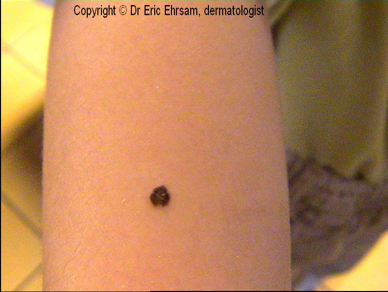
A 67-year old woman with immunosuppression (chemotherapy for a disseminated breast cancer) consulted for a pruritic rash with papulovesicular lesions and squamous lesions on her hands.


The clinical diagnosis was a Norwegian scabies. Many nurses of the department developed a scabies during the following weeks after the entrance of this patient.


Dermoscopy of lesions on her hands revealed a brown triangle (arrows) corresponding
to the head of the mite. If the mite is seen from the dorsal, the triangle appears angulated (see the above picture) or not angulated (►) if it is seen from the ventral site.


On this dermoscopic picture taken from the palmar face, many scales and mites can be seen.

Friday 7 December 2007

Melanocytic nevus


A 28-year-old woman consulted for an enlarging scapular nevus on her lefts scapular area.
The presence of an irregular depigmentation and blue-gray dots (circle) in favor of a regression lead to excise it. Pathology revealed a benign dermal melanocytic nevus.


Wednesday 5 December 2007

Basal cell carcinoma


A 69-year-old patient presented a pigmented tumor on his right temporal area.
Dermoscopy revealed blue ovoid nests in favour of a basal cell carcinoma.
The lesion was removed and pathology confirmed the dermoscopic diagnosis.

Parallel furrow pattern


A 18-year-old man consulted for 3 palmar nevi. Dermoscopy showed typical parallel furrow patterns in favour of benign acral nevi


Tuesday 4 December 2007

Unna nevus


Melanocytic nevus on the abdomen of a 35-year-old woman.
Dermoscopy reveals exophytic papillary structures, comedo-like openings, milia-like cysts and comma-like vessels.


Monday 3 December 2007

Rim of globules


A 16-year-old boy consulted for his nevi after summer holidays. Dermoscopy revealed 3 nevi with rim of globules. This dermoscopic feature is often seen after sun exposition and in enlarging nevi.


Spitz nevus


11-year-old boy with a dark pigmented lesion on his right arm.
Dermoscopy: symetric starburst pattern, streaks, regular globules at the center of the lesion. The diagnosis of Spitz nevus was made and a dermoscopic follow-up did not reveal enlargement or changing of structures.