Search This Blog

Showing posts with label peppering. Show all posts

Wednesday, 4 June 2014

Regression


A 48-year-old woman presented this pigmented lesion on her back. 


 1: clinical picture

 2: dermoscopic picture

3: dermoscopic picture

  4:  dermoscopic picture


Dermoscopy revealed:
  • regression areas with some depigmentation (green arrows and ovoid area)
  • blue gray structures (blue arrows) and blue gray dots (peppering)
  • linear irregular vessels (black arrows)


Lesion was excised and pathology revealed a regressive superficial spreading melanoma with Breslow index at  0,39 mm

Friday, 22 July 2011

Back lesion


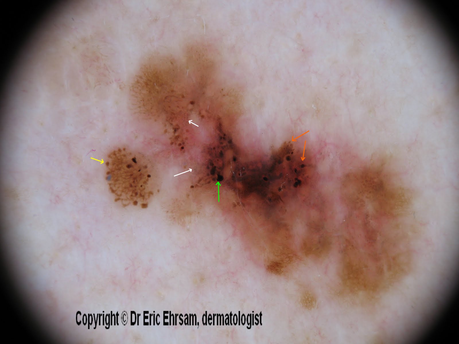
A 54-year-old woman consulted for this changing lesion on her back.


Dermoscopy revealed:

  • multiple colours, irregular pigmentation
  • a multi-component pattern
  • an atypical reticular pattern
  • irregular streaks (yellow arrow)
  • irregular dots and globules (orange arrow)
  • irregular blotches (green arrow)
  • peppering, regression (white arrow)


Pathology revealed a superficial spreading melanoma (SSM) with a Breslow index at 0.33 mm

Thursday, 21 October 2010

Regression


A 61-year-old man consulted for a pigmented lesion on his back.

Clinically this lesion was asymmetrical with 2 colours: pink and black.

Dermoscopy revealed: regression signs with a pink area, blue gray dots with a peppering aspect.

Pathology report: fibrosis and inflammation, hypermelanosis. Melanophages developed on a 1 mm depth. Immunochemistry: anti-human A negative.

In conclusion, this lesion was a fully regressive melanoma.


We recommend to read this article:

Dermoscopy of fully regressive melanoma. L. Thomas and al.
The British Journal of Dermatology. 2008;158(6):1224-1229

Wednesday, 14 October 2009

Superficial spreading melanoma on an arm


A 31-year-old man consulted for this pigmented lesion on his right arm.


Dermoscopy revealed pigmented lesion with the following dermoscopic signs:
  • asymmetry of colors and shape
  • regression area: depigmentation (black rectangle ) and blue gray dots (peppering) (yellow circle)
  • atypical reticular network (brown rectangle)
  • pseudopods (red arrow)

This lesion was excised and pathology confirmed the diagnosis of superficial spreading melanoma. Breslow index was 0,48 mm.

Saturday, 10 May 2008

Peppering


A 85-year-old woman consulted for a slowly enlarging pigmented lesion on her right cheek


Dermoscopy revealed a pseudonetwork which is characteristic of facial pigmented lesions.
Furthermore, dermoscopy showed multiple slate-grays dots or pepper-like granules realizing a typical an annular-granular pattern or peppering in favor of a lentigo maligna melanoma. A biopsy confirmed this diagnosis.


Peperring

Peppering or annular-granular pattern can also be present in lichen planus-like keratosis and actinic keratosis.
Some lentigo maligna melanomas exhibit this structure.

Sunday, 18 November 2007

Eccentric pigmentation

This lesion presented an asymmetric reticular network with an eccentric pigmentation, globules, and signs of regression with blue-gray dots (peppering).
The lesion was excised and pathology revealed a junctional melanocytic nevus


Tuesday, 18 September 2007

Melanoma with regression

A 62-year-old patient with a superficial spreading melanoma on his right shoulder.


Dermoscopy (x20) revealed white areas (scar-like depigmentation) and delicate peppering.


Other cases of melanoma
0 1 2 3 4 5 6 7 8 9 10 11 12 13 14 15 16 17 18

Friday, 14 September 2007

Superficial spreading melanoma


A 61-year-old man consulted for this lesion located on his trunk (lateral left side).


Dermoscopy revealed an atypical reticular pattern, with thickened and broken net, regression structures, with blue pepper-like granules.
The lesion was removed and pathology revealed a superficial spreading melanoma (SSM) with Breslow index mesured at 0,6mm.
No metastases were detected.


Other cases of melanoma
0 1 2 3 4 5 6 7 8 9 10 11 12 13 14 15 16 17 18