Search This Blog

Showing posts with label blue gray dot. Show all posts

Saturday, 28 November 2015

Regression.


A 21-year-man consulted for this lesion on his arm


Dermoscopy revealed:

  • a whitish - pinkish structureless area
  • with gray dots
in favor of a regressive lesion.

Pathology was in favor of a fully regressive melanocytic lesion. A fully regressive melanoma cannot be ruled out. 

Wednesday, 4 June 2014

Regression


A 48-year-old woman presented this pigmented lesion on her back. 


 1: clinical picture

 2: dermoscopic picture

3: dermoscopic picture

  4:  dermoscopic picture


Dermoscopy revealed:
  • regression areas with some depigmentation (green arrows and ovoid area)
  • blue gray structures (blue arrows) and blue gray dots (peppering)
  • linear irregular vessels (black arrows)


Lesion was excised and pathology revealed a regressive superficial spreading melanoma with Breslow index at  0,39 mm

Friday, 23 March 2012

Red lesion on an arm...


 A 50-year-old woman consulted for an enlarging lesion on her right arm;
Clinical examination revealed a red papular lesion with a slight pigmentation.


Dermoscopy revealed:

  •  atypical vascular pattern with polymorphous vessels, namely linear irregular vessels, dotted vessels
  • a small area of pigmentation (gray dots)
An excision was performed and pathology revealed an achromic melanoma with a Breslow index at 0.5mm.

Thursday, 21 October 2010

Regression


A 61-year-old man consulted for a pigmented lesion on his back.

Clinically this lesion was asymmetrical with 2 colours: pink and black.

Dermoscopy revealed: regression signs with a pink area, blue gray dots with a peppering aspect.

Pathology report: fibrosis and inflammation, hypermelanosis. Melanophages developed on a 1 mm depth. Immunochemistry: anti-human A negative.

In conclusion, this lesion was a fully regressive melanoma.


We recommend to read this article:

Dermoscopy of fully regressive melanoma. L. Thomas and al.
The British Journal of Dermatology. 2008;158(6):1224-1229

Wednesday, 14 October 2009

Superficial spreading melanoma on an arm


A 31-year-old man consulted for this pigmented lesion on his right arm.


Dermoscopy revealed pigmented lesion with the following dermoscopic signs:
  • asymmetry of colors and shape
  • regression area: depigmentation (black rectangle ) and blue gray dots (peppering) (yellow circle)
  • atypical reticular network (brown rectangle)
  • pseudopods (red arrow)

This lesion was excised and pathology confirmed the diagnosis of superficial spreading melanoma. Breslow index was 0,48 mm.

Tuesday, 13 October 2009

Facial lesion


A 55-year-old woman consulted for this pigmented lesion on her forehead.
This lesion was present for 3 years and had progressively changed of colour.


Dermoscopy revealed:
  • multiple colours
  • an atypical reticular network (white square)
  • regression areas: depigmentation and blue grey dots (yellow square)
  • irregular blotches (white circle)
  • follicular plugs (red arrow)
This lesion was excised and pathology revealed a superficial spreading melanoma with a Breslow Index at 0.24mm.

Thursday, 26 March 2009

melanoma

A 82-year-old woman consulted for this dark pigmented lesion on her left arm. Clinically a melanoma was suspected.
Dermoscopy revealed:
  • a multi-coloured pattern
  • a blue white veil
  • blue gray dots
  • whitish scar-like depigmentation
  • polymorphous vessels including linear irregular vessels


Monday, 16 March 2009

scalp melanoma


A 65-year-old man consulted for this pigmented lesion on his scalp.

Dermoscopy revealed a disorganised atypical reticular network, blue gray dots and regression, irregular branched streaks.
This lesion was excised and pathology revealed a melanoma in situ.


Saturday, 21 June 2008

Basal cell carcinoma

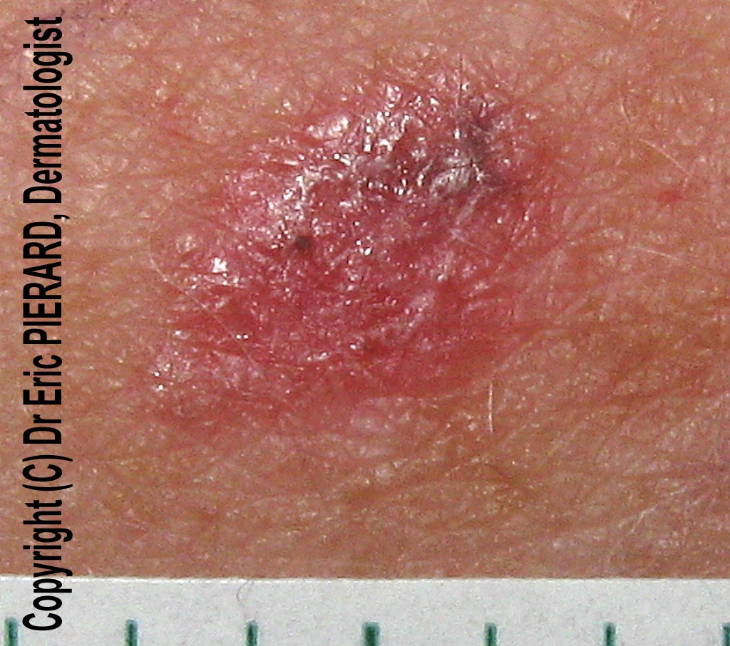
A 89-year-old woman consulted for a pigmented lesion on her forehead. A basal cell carcinoma was the main clinical diagnosis.

On this picture, with a slight pressure of the dermoscope (polarized light dermoscopy), arborizing blood vessels are well seen.
Blue gray dots and globules are the other dermoscopic signs in favor of this basal cell carcinoma.

If the pressure of the dermoscope is too important, the blood vessels are not visible.

Vessels and red areas are better visualized with polarized light dermoscopy than with immersion contact dermoscopy (1)

References:
1 - Marghoob et al. Differences between polarized light dermoscopy and immersion contact dermoscopy for the evaluation of skin lesions. Arch Dermatol 2007 Mar;143(3):329-38


Friday, 7 December 2007

Melanocytic nevus


A 28-year-old woman consulted for an enlarging scapular nevus on her lefts scapular area.
The presence of an irregular depigmentation and blue-gray dots (circle) in favor of a regression lead to excise it. Pathology revealed a benign dermal melanocytic nevus.


Friday, 30 November 2007

Seborrheic keratosis


A 90-year-old woman consulted for a pigmented lesion on her forehead.
Dermoscopy revealed a brown pigmentation with multiple blue-gray dots.
The lesion was excised and pathology revealed a seborrheic keratosis.

This lesion was probably regressing to a lichenoïd keratosis (or lichen planus-like keratosis).


See the literature:

Zaballos P. et al. Dermoscopic pattern of intermediate stage in seborrhoeic keratosis regressing to lichenoid keratosis: report of 24 cases. Br J Dermatol. 2007 Aug;157(2):266-72

Sunday, 18 November 2007

Eccentric pigmentation

This lesion presented an asymmetric reticular network with an eccentric pigmentation, globules, and signs of regression with blue-gray dots (peppering).
The lesion was excised and pathology revealed a junctional melanocytic nevus


Saturday, 17 November 2007

Regression structures

Asymmetrical multicoloured pattern with regression structures:
- blue-white veil
- scar-like depigmentation
and blue-gray dots
The diagnosis was a superficial spreading melanoma, Breslow thickness 0.55mm.


Other cases of melanoma
0 1 2 3 4 5 6 7 8 9 10 11 12 13 14 15 16 17 18