Search This Blog

Showing posts with label dots. Show all posts

Saturday, 28 November 2015

Regression.


A 21-year-man consulted for this lesion on his arm


Dermoscopy revealed:

  • a whitish - pinkish structureless area
  • with gray dots
in favor of a regressive lesion.

Pathology was in favor of a fully regressive melanocytic lesion. A fully regressive melanoma cannot be ruled out. 

Pigmented lesion on neck.


A 57 year-old-woman consulted for a pigmented lesion on her neck.


Dermoscopy revealed:
  • no melanocytic structures
  • a structureless pattern
  • irregular blue gray dots/globules 


Pathology was in favor of a Bowen's disease.

Friday, 22 July 2011

Back lesion


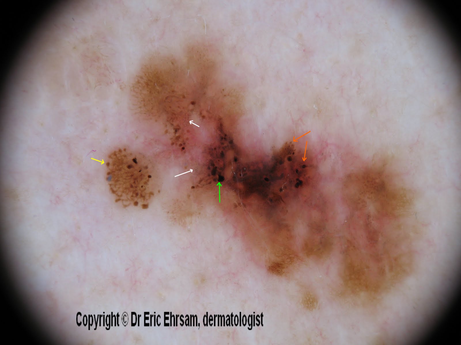
A 54-year-old woman consulted for this changing lesion on her back.


Dermoscopy revealed:

  • multiple colours, irregular pigmentation
  • a multi-component pattern
  • an atypical reticular pattern
  • irregular streaks (yellow arrow)
  • irregular dots and globules (orange arrow)
  • irregular blotches (green arrow)
  • peppering, regression (white arrow)


Pathology revealed a superficial spreading melanoma (SSM) with a Breslow index at 0.33 mm

Tuesday, 24 August 2010

Pigmented breast lesion

A 28-year-old woman consulted for an enlarging pigmented lesion on her left breast.

Dermoscopy revealed:
  • a multi-colored pattern
  • a multicomponent pattern
  • asymmetry in 2 axes
  • an atypical reticular pattern
  • some atypical dots/globules
  • a scar-like depigmentation
  • a blue white veil
Pathology revealed a superficial spreading melanoma with a Breslow index at 0,5mm

Wednesday, 6 August 2008

Superficial spreading melanoma


A 66-year-old man consulted for a pigmented lesion on his left scapular area.

Dermoscopy revealed a multi-component pattern, asymmetry, and multiple colours (tan, dark brown, black, blue gray, white).
Other signs were an atypical reticular pattern (irregular holes and thick lines) with a sharp demarcation, a blue-white veil and atypical vessels (irregular linear vessels), a central ulceration (crust). Dots were irregularly distributed.

All these dermoscopic signs were in favor of a superficial spreading melanoma.

Monday, 17 March 2008

Acral melanocytic nevus

A 6-year-old boy consulted for a congenital acral melanocytic nevus on his 2nd left toe.


This lesion exhibited a combination of 3 patterns, namely a parallel furrow pattern (dots on the furrows), an homogeneous pattern and a reticular pattern.


Parallel furrow pattern (circle) and reticular pattern (rectangle) were the main patterns observed on this benign melanocytic nevus.

Other cases of acral nevi:
1  2  3  4  5  6  7  8  9  10  11  

Early superficial spreading melanoma

A 37-year-old man consulted for an enlarging pigmented lesion on his abdomen.


Dermoscopy revealed an atypical pattern with an asymmetry, homogeneous area, irregular dots and globules, and irregular branched streaks.


The lesion was excised and pathology revealed a superficial spreading melanoma with a Breslow index non measurable.

Other cases of melanoma
0 1 2 3 4 5 6 7 8 9 10 11 12 13 14 15 16 17 18

Wednesday, 28 November 2007

Lentigo maligna melanoma


A 58-year-old woman consulted for this non homogeneous lesion on her left forearm.


Dermoscopy revealed a lightly pigmented network that was focally darker on certain areas or absent of other areas, and the presence of rare brown dots.
The red area was due to a punch biopsy.


A biopsy was performed and revealed a lentigo maligna melanoma. The lesion was completely excised and pathology revealed LMM with a Breslow index at 0.10mm


Sunday, 25 November 2007

Atypical (dysplastic) nevus

A 68-year-old woman consulted for this pigmented lesion on her back.
Dermoscopy revealed an asymmetric lesion with an atypical reticular and homogenous pattern , a few subtle blue-white structures (circle) and brown dots (arrow)
The lesion was excised and pathology revealed an atypical (dysplastic) compound type.


Tuesday, 6 November 2007

Pseudopods

A 60-year-old man presented a nodular pigmented tumor on his chest. Dermoscopy revealed a pigment network at the periphery with many pseudopods (white circle), irregular dots and globules at 6 o'clock and 3 o'clock, and a blue white veil. The lesion was excised and pathology revealed superficial spreading melanoma with a Breslow index at 1.5mm and a Clark level II.

Other cases of melanoma
0 1 2 3 4 5 6 7 8 9 10 11 12 13 14 15 16 17 18

Tuesday, 9 October 2007

Atypical congenital nevus


A 14 weeks-old child presented an atypical congenital nevus on her left arm.

Dermoscopy showed an asymetric lesion with blue white veil and irregular dots at the periphery.

The lesion was excised and pathology revealed a benign congenital compound nevus.

Monday, 13 August 2007

superficial spreading melanoma


This case concerned a 59-year-old patient: this pigmented lesion of his right thigh was discovered during a consultation for condylomas. The patient was not conscious of this lesion.

On dermoscopy (x 20) : Multicomponent pattern, irregular pigment network, blue white veil very irregular, irregular streaks (bottom) irregular dots and globules.


Other cases of melanoma
0 1 2 3 4 5 6 7 8 9 10 11 12 13 14 15 16 17 18

Saturday, 11 August 2007

superficial spreading melanoma


A 84-year-old man presenting many seborrheic keratoses, developed a slowly enlarging pigmented lesion of the abbdomen


On dermoscopy (x20): asymetric lesion with atypical globular pattern, irregular dots and globules at 3 o clock (right side) , and blue white veil


The lesion was removed and pathologist concluded to a superficial spreading melanoma Breslow index 0.28mm


Other cases of melanoma
0 1 2 3 4 5 6 7 8 9 10 11 12 13 14 15 16 17 18