Search This Blog

Showing posts with label melanoma (superficial spreading). Show all posts

Wednesday, 11 May 2016

Eccentric pigmentation


36-year-old woman with an atypical pigmented lesion on her left leg


Demoscopy revealed:

  • an eccentric hyperpigmentation
  • an atypical reticular network
  • pseudopods


Pathology revealed a superficial spreading melanoma with Breslow index at 0.25 mm

Friday, 12 February 2016

Brachial melanoma


A 54-year-old man had a rapidly growing lesion on his right back for 6 months.


Dermoscopy revealed:
  • a vascular pattern with some polymorphous and atypical vessels
  • peripheral pigmentation
  • irregular globules
  • eccentric blotches


Pathology revealed a superficial spreading melanoma with Breslow index at 1.53 mm

Saturday, 8 November 2014

Starburst pattern


A 43-year-old man consulted for this acquired pigmented lesion on his left leg.


Dermoscopy revealed:
  • a central blotch
  • a regular starburst pattern


 Two diagnoses were evoked: Reed nevus or melanoma. Regular blotch and symmetrical starburst pattern favored Reed nevus.

Lesion was excised and pathology revealed a superficial spreading melanoma with a Breslow index at 0.28mm.

Saturday, 4 October 2014

An ugly duckling


A 26-year-old woman consulted for many moles. One of them was considered "an ugly duckling".


Dermoscopy revealed:
  • an irregular blotch
  • asymmetry of form
  • chrysalis-like structures
Pathology revealed a superficial spreading melanoma (SSM) with a Breslow index at 0,6mm

Sunday, 7 September 2014


A 32-year-old man consulted for this enlarging pigmented lesion.


Clinical presentation: multiple couleurs, asymmetry

Dermoscopy revealed:

  • multiple colors
  • irregular dots
  • pink structureless area
  • irregular blotch
  • regression
This lesion was easily diagnosed superficial spreading melanoma.

Pathology revealed a superficial spreading melanoma with a Breslow index at 0.5mm

Wednesday, 4 June 2014

Regression


A 48-year-old woman presented this pigmented lesion on her back. 


 1: clinical picture

 2: dermoscopic picture

3: dermoscopic picture

  4:  dermoscopic picture


Dermoscopy revealed:
  • regression areas with some depigmentation (green arrows and ovoid area)
  • blue gray structures (blue arrows) and blue gray dots (peppering)
  • linear irregular vessels (black arrows)


Lesion was excised and pathology revealed a regressive superficial spreading melanoma with Breslow index at  0,39 mm

Wednesday, 12 March 2014


A 41-year-old woman was examined for this lesion on her right thigh.


Dermoscopy revealed:

  • a multicomponent pattern
  • a reticular depigmentation  
  • chrysalis-like structures
  • polymorphous vessels including corkscrew vessels
Pathology revealed a superficial spreading melanoma with a Breslow index at 0.54mm

Monday, 31 December 2012

A pigmented lesion on back


A 75-year-old man consulted for this pigmented lesion on his back.
His skin was heavily sun exposed.
Clinical image is in the lower right corner.
Dermoscopy revealed:
- an atypical reticular network with different colours and disorganization
- a blue structureless area

This lesion was excised and pathology revealed a superficial spreading melanoma.
Breslow index was 0,30 mm.

Friday, 22 July 2011

Back lesion


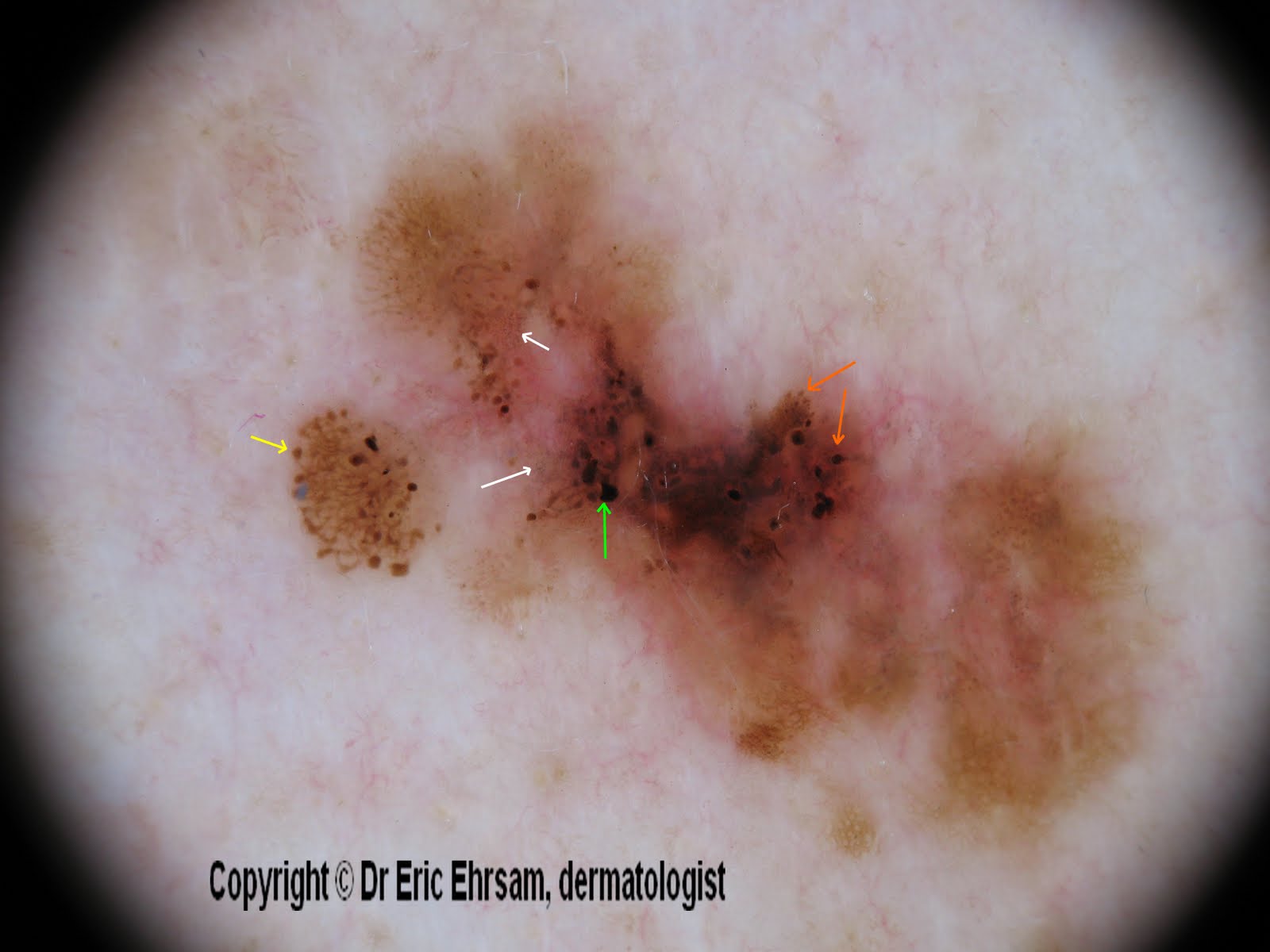
A 54-year-old woman consulted for this changing lesion on her back.


Dermoscopy revealed:

  • multiple colours, irregular pigmentation
  • a multi-component pattern
  • an atypical reticular pattern
  • irregular streaks (yellow arrow)
  • irregular dots and globules (orange arrow)
  • irregular blotches (green arrow)
  • peppering, regression (white arrow)


Pathology revealed a superficial spreading melanoma (SSM) with a Breslow index at 0.33 mm

Monday, 23 May 2011

Emerging on a forearm


A 73-year-old man consulted for a pigmented lesion on his right forearm. This lesion was not symmetrical and been recently discovered by patient.


Dermoscopy revealed:
  • an asymmetrical pigmentation
  • a disorganised and atypical reticular network
  • an homogeneous area
This lesion was excised and pathology revealed a superficial spreading melanoma in situ. 

Saturday, 7 May 2011

Forearm lesion


A 59-year-old woman consulted for this pigmented lesion on her right forearm.


Dermoscopy revealed:
  • irregular pigmentation
  • atypical reticular pattern
  • irregular dots / globules
  • brown dots
  • regressive homogeneous area
in favor of a melanoma.


This lesion was excised and pathology revealed a superficial spreading melanoma with a Breslow index at 0,47mm

Tuesday, 24 August 2010

Pigmented breast lesion

A 28-year-old woman consulted for an enlarging pigmented lesion on her left breast.

Dermoscopy revealed:
  • a multi-colored pattern
  • a multicomponent pattern
  • asymmetry in 2 axes
  • an atypical reticular pattern
  • some atypical dots/globules
  • a scar-like depigmentation
  • a blue white veil
Pathology revealed a superficial spreading melanoma with a Breslow index at 0,5mm

Wednesday, 18 August 2010

What is this lesion on a leg (2nd case) ?


A 71-year old woman consulted for this enlarging multi-colored lesion on her right leg.

Dermoscopy revealed:
  • irregular pigmentation
  • atypical pigment network
  • negative pigment network
  • irregularly distributed blotch
  • blue-white veil
in favor of a melanoma.

An excision was performed and pathology revealed a superficial spreading melanoma in vertical extension, Clark level IV, and Breslow index at 3,1 mm.

Wednesday, 28 July 2010

Acquired pigmented lesion on a forearm

A 62-year-old woman consulted for an enlarging lesion on her right forearm.

Dermoscopy revealed an asymmetric reticular pattern with irregular dots.

Pathology revealed a superficial spreading melanoma with a Breslow index at 0.23mm.

Saturday, 24 July 2010

A changing lesion on his back...

A 30-year-old man consulted for a pigmentend lesion on his back. His wife noticed this lesion was slowly changing.

Dermoscopy revealed an atypical a reticular network, an irregular pigmentation, and an atypical vascular pattern.

This lesion was excised and pathology revealed a superficial spreading melanoma with a vertical extension, with a Breslow index at 1.3mm.

Tuesday, 13 July 2010

Lost among black benign lesions....


A 72-year-old patient consulted for many pigmented lesions on his back. Most of them were seborrheic keratoses...........

............except this one.

Dermoscopy revealed:
  • a multi- colored pattern
  • a sharp demarcation
  • an atypical reticular network
  • a blue white veil
This lesion was excised and pathology revealed a superficial spreading melanoma (Breslow index 0.72mm) developed on a melanocytic nevus

Wednesday, 14 October 2009

Superficial spreading melanoma on an arm


A 31-year-old man consulted for this pigmented lesion on his right arm.


Dermoscopy revealed pigmented lesion with the following dermoscopic signs:
  • asymmetry of colors and shape
  • regression area: depigmentation (black rectangle ) and blue gray dots (peppering) (yellow circle)
  • atypical reticular network (brown rectangle)
  • pseudopods (red arrow)

This lesion was excised and pathology confirmed the diagnosis of superficial spreading melanoma. Breslow index was 0,48 mm.

Tuesday, 13 October 2009

Facial lesion


A 55-year-old woman consulted for this pigmented lesion on her forehead.
This lesion was present for 3 years and had progressively changed of colour.


Dermoscopy revealed:
  • multiple colours
  • an atypical reticular network (white square)
  • regression areas: depigmentation and blue grey dots (yellow square)
  • irregular blotches (white circle)
  • follicular plugs (red arrow)
This lesion was excised and pathology revealed a superficial spreading melanoma with a Breslow Index at 0.24mm.

Thursday, 2 July 2009

Blue white veil


A 72-year-old man consulted for this black tumour on his back.


Dermoscopy revealed a sharply multi-coloured lesion without visible reticular pigment network covered by a central crust.


Most strikingly features were:
  • a blue white veil
  • irregular and thick vessels (see white square)
in favour of a melanoma.