Search This Blog

Showing posts with label multiple colours. Show all posts

Friday, 27 November 2015

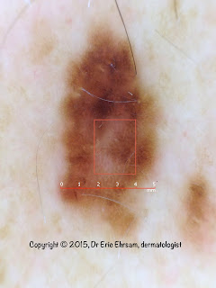
Back lesion


A 65-year-old man with many pigmented lesions on his back. One of this lesion was atypical.


Dermoscopy revealed:
  • multiple colors
  • an atypical reticular network
  • regression
  • a reticular depigmentation  (rectangle)


Pathology revealed a melanoma in situ.

Sunday, 7 September 2014


A 32-year-old man consulted for this enlarging pigmented lesion.


Clinical presentation: multiple couleurs, asymmetry

Dermoscopy revealed:

  • multiple colors
  • irregular dots
  • pink structureless area
  • irregular blotch
  • regression
This lesion was easily diagnosed superficial spreading melanoma.

Pathology revealed a superficial spreading melanoma with a Breslow index at 0.5mm

Friday, 12 July 2013

A slowly enlarging pigmented lesion


A 32-year-old woman consulted for a pigmented lesion on her left arm. This lesion was slowly enlarging for 3 years.

 Dermoscopy revealed:

  • an atypical reticular pattern with a total disorganization
  • multiple colors
  • signs of regression


 Two punch biopsies were performed.
Pathology was in favor of melanoma in situ.
Lesion was excised with a 0.5 margin.


Tuesday, 23 August 2011

Pectoral lesion


A 52-year-old man presented this pectoral lesion. No clinical history was available.


Dermoscopy revealed:

  • multiple colours
  • a multi-component pattern
  • an atypical reticular pattern
  • some pseudopods 
  • irregular blotches
  • regression areas
Pathology confirmed the diagnosis of superficial spreading melanoma.

Tuesday, 24 August 2010

Pigmented breast lesion

A 28-year-old woman consulted for an enlarging pigmented lesion on her left breast.

Dermoscopy revealed:
  • a multi-colored pattern
  • a multicomponent pattern
  • asymmetry in 2 axes
  • an atypical reticular pattern
  • some atypical dots/globules
  • a scar-like depigmentation
  • a blue white veil
Pathology revealed a superficial spreading melanoma with a Breslow index at 0,5mm

Tuesday, 13 October 2009

Facial lesion


A 55-year-old woman consulted for this pigmented lesion on her forehead.
This lesion was present for 3 years and had progressively changed of colour.


Dermoscopy revealed:
  • multiple colours
  • an atypical reticular network (white square)
  • regression areas: depigmentation and blue grey dots (yellow square)
  • irregular blotches (white circle)
  • follicular plugs (red arrow)
This lesion was excised and pathology revealed a superficial spreading melanoma with a Breslow Index at 0.24mm.

Thursday, 26 March 2009

melanoma

A 82-year-old woman consulted for this dark pigmented lesion on her left arm. Clinically a melanoma was suspected.
Dermoscopy revealed:
  • a multi-coloured pattern
  • a blue white veil
  • blue gray dots
  • whitish scar-like depigmentation
  • polymorphous vessels including linear irregular vessels


Wednesday, 6 August 2008

Superficial spreading melanoma


A 66-year-old man consulted for a pigmented lesion on his left scapular area.

Dermoscopy revealed a multi-component pattern, asymmetry, and multiple colours (tan, dark brown, black, blue gray, white).
Other signs were an atypical reticular pattern (irregular holes and thick lines) with a sharp demarcation, a blue-white veil and atypical vessels (irregular linear vessels), a central ulceration (crust). Dots were irregularly distributed.

All these dermoscopic signs were in favor of a superficial spreading melanoma.

Saturday, 1 December 2007

Superficial spreading melanoma


A 50-year-old man presented an irregularly pigmented lesion his back. Dermoscopy revealed an asymetric pigmentation, multiple colors, streaks (red circle), and a delicate blue white veil in favor of a superficial spreading melanoma.
The lesion was removed with a 2 mm margin and histology confirmed the diagnosis. The Breslow index was measured 0.63 mm. A re-excision was performed with a 1 cm margin. No mestastases were detected.


Other cases of melanoma
0 1 2 3 4 5 6 7 8 9 10 11 12 13 14 15 16 17 18

Tuesday, 27 November 2007

Invasive melanoma


A 62-year-old man consulted for this multicolored tumor on his scapular area. This lesion was clinically in favor of an advanced invasive melanoma (nodular melanoma).


Asymmetrical pigmentation pattern, multiple colours , atypical vessels, atypical reticular pattern, blue-white veil, and ulceration were found in this case of invasive melanoma, Breslow thickness 9mm


Other cases of melanoma
0 1 2 3 4 5 6 7 8 9 10 11 12 13 14 15 16 17 18

Friday, 23 November 2007

Superficial spreading melanoma

The clinical aspect of this pigmented lesion was in favor of a superficial spreading melanoma.
Dermoscopy showed multiple colors (tan, dark brown, black, gray, blue), areas of regression (scar-like depigmentation), a blue white veil.
The lesion was excised and pathology revealed a superficial spreading melanoma, Breslow thickness 0.56mm.


Other cases of melanoma
0 1 2 3 4 5 6 7 8 9 10 11 12 13 14 15 16 17 18