Search This Blog
Showing posts with label homogeneous pattern. Show all posts
Showing posts with label homogeneous pattern. Show all posts
Sunday, 21 April 2013
A 60-year-old man consulted for this black-blue tumor on his back.
Patient did not know when this lesion appeared.
Dermoscopy revealed a homogenous blue pattern with whiter areas.
Two diagnosis were suspected:
- blue nevus
- melanoma
This lesion was excised and pathology revealed a blue nevus with fibrous involution
Saturday, 1 September 2012
A new melanoma ?
A 30-year-old woman consulted for this black pigmented lesion on her 4th right toe. She mentioned to have tried to remove it with a blade and a needle.
Dermoscopy revealed a black homogeneous pattern.
A melanoma was suspected and lesion was surgically removed for pathology.
Pathology revealed a wart.
This is a quite unusual presentation for a wart.
Libellés :
black pigmentation,
homogeneous pattern,
wart
Wednesday, 3 February 2010
Blue lesion on a leg
 A 58-year-old woman consulted for a pigmented lesion on her right leg present for many years.
NB: a scar is due to a former fracture.
Libellés :
Blue nevus,
homogeneous blue pattern,
homogeneous pattern
Wednesday, 18 March 2009
Actinic lentigo

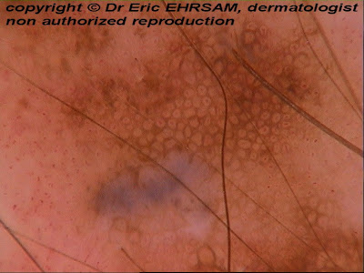
A 72-year-old man consulted for an enlarging pigmented lesion on his right hand. This lesion was quite typical of an actinic lentigo.
Dermoscopy confirmed easily this diagnosis showing: a homogeneous pattern and a pseudoreticular network with thin lines, with a moth-eaten border

Libellés :
homogeneous pattern,
lentigo,
moth-eaten border,
pseudo-network
Friday, 18 April 2008
Atypical (dysplastic) nevi
 A 19-year-old man consulted for multiple atypical (dysplastic nevi or Clark nevi) on her trunk. Three of them were clustered on her back. They featured three different dermoscopic patterns:
1-reticulo-globular pattern with eccentric hyperpigmentation

Saturday, 5 April 2008
Combined nevus
 A 16-year-old girl consulted for a melanocytic nevus with 2 colors located on her back.
Dermoscopy revealed a globular pattern associated with a central homogenous pattern (darker area) in favor of a benign combined nevus.

A combined nevus is a nevus which contains two or more components, most often with 2 distinct colors.
In some cases combined nevus consists of a typical blue nevus associated with another kind of nevus.
A combined nevi can clinically mimick melanomas because of the markedly variegated coloration and asymmetry suggestive of a melanoma occuring on pre-existing nevus.
Dermoscopically , combined nevi present with a multicomponent pattern (homogenous, reticular and globular in combinations )
If a combined nevus is composed of a blue nevus and a common nevus, dermoscopy reveals the stereotypical findings of a blue nevus, namely ( homogenous blue-gray pigmentation without observable local features)
Tuesday, 25 March 2008
Blue nevus

A 43-year-old woman consulted for a dark bue lesion on her left buttock.
 Dermoscopy revealed an homogeneous pattern made of bluish to white-blue structureless pigmentation, in favor of a blue nevus.
Libellés :
Blue nevus,
homogeneous pattern,
structureless area
Monday, 17 March 2008
Acral melanocytic nevus
 A 6-year-old boy consulted for a congenital acral melanocytic nevus on his 2nd left toe.
This lesion exhibited a combination of 3 patterns, namely a parallel furrow pattern (dots on the furrows), an homogeneous pattern and a reticular pattern.

Parallel furrow pattern (circle) and reticular pattern (rectangle) were the main patterns observed on this benign melanocytic nevus.
Wednesday, 2 January 2008
Compound melanocytic nevus

A 14-year-old girl consulted for an enlarging melanocytic nevus on her back. One of her sister, aged 24, had been operated for a superficial spreading melanoma 2 years before, and she was very anxious. The lesion was larger than 6 mm and had an irregular border.

Dermoscopy revealed a central homogeneous pattern and a peripheral typical reticular pattern, and peripheral globules, but no signs for a SSM.
The lesion was removed and pathology was in favour of a compound melanocytic nevus.
Monday, 26 November 2007
Acral melanocytic nevus


A 39-year-old woman (phototype V) noticed a pigmented lesion on her 5th left toe.
Dermoscopy revealed a uniform black homogeneous pattern with whitish dots (eccrine pores) and some streaks.
The lesion was excised and pathology revealed a compound melanocytic nevus.
Sunday, 25 November 2007
Atypical (dysplastic) nevus
 A 68-year-old woman consulted for this pigmented lesion on her back.
Dermoscopy revealed an asymmetric lesion with an atypical reticular and homogenous pattern , a few subtle blue-white structures (circle) and brown dots (arrow)
 The lesion was excised and pathology revealed an atypical (dysplastic) compound type.

Saturday, 17 November 2007
Spitz nevus
 A 14-year-old boy presented a dark pigmented lesion on his lumbar area. Dermoscopy revealed a homogeneous pattern with a bluish pigmentation. Rare brown globules were found at the periphery. The lesion was excised and pathology revealed a dermal Spitz nevus.
Six dermoscopic patterns of Spitz nevus have been described:
-vascular pattern with dotted vessels
-globular pattern
-reticular pattern
-starburst pattern
-homogeneous pattern
-atypical pattern
Libellés :
globule,
homogeneous pattern,
melanocytic nevus (Spitz)
Tuesday, 23 October 2007
Blue nevus
 A 32-year-old woman consulted for this blue lesion on her nose.
Dermoscopy revealed an homogeneous blue pattern in favor of a blue nevus.


Tuesday, 14 August 2007
Atypical nevus
 A 40-year-old man consulted for this atypical mole of the intermammary region, without recent changing.
 Dermoscopy (x20) showed a combination of two patterns:
1-on the left: an homogenous pattern with comma vessels
2- on the right: a reticular pattern with blue white veil
The lesion was removed and pathology revealed a benign irritated compound nevus with lateral junctionnal extension
Subscribe to:
Posts (Atom)


