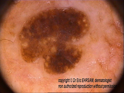
A 53-year-old woman consulted for this pigmented lesion on her back.
Dermoscopy revealed brown structures typical of fat fingers, in favor of a seborrheic keratosis.
Fat fingers are an important dermoscopic sign to know for diagnosing seborrheic keratosis in absence of other typical signs (milia-like kysts, comedo-like structures, fingerprint-like structures, sharp margins, hairpin blood vessels)
Kopf and al, "Fat fingers", a clue in the dermoscopic diagnosis of seborrheic keratoses J Am Acad Dermatol
2006;55:1089-91
Search This Blog
Monday, 29 October 2007
Saturday, 27 October 2007
Squamous cell carcinoma
A 78-year-old man consulted for this rapidly enlarging tumor on the left side of his nose. The lesion was present for 3 months. The possible clinical diagnoses were keratoacanthoma, squamous cell carcinoma, and basal cell carcinoma.
Dermoscopy revealed whitish halo (A) and linear-irregular vessels (B). The lesion was excised and pathology was in favor of a squamous cell carcinoma.
Libellés :
linear-irregular vessel,
squamous cell carcinoma,
white halo
Seborrheic keratosis
A 50-year-old woman presented a pigmented tumor on her left arm.
Dermoscopy revealed many comedo-like openings, crypts, and milia-like cysts inf favor of a seborrheic keratosis.
Libellés :
comedo-like opening,
crypts,
milia-like cyst,
seborrheic keratosis
Friday, 26 October 2007
Atypical dermatofibroma with vascular pattern
A 68-year-old man consulted for this lesion on his right leg present for more than 15 years.
Dermoscopy revealed some light brown globules (*) and a vascular pattern with polymorphous vessels: hairpin vessels, dotted vessels, and linear-irregular vessels.
A biopsy was performed and revealed a dermatofibroma.
Dermatofibromas with vascular pattern have been reported in the literature (D.Picolo, K.Peris, page 287, in color atlas of melanocytic lesions of the skin, Springer edition, 2007)
Dermatofibroma
A 30-year-old woman consulted for this pigmented lesion on her right leg.
Dermoscopy revealed a central white scar-like patch surrounded by a delicate pigmented network, in favor of a dermatofibroma.
Libellés :
central white patch,
Dermatofibroma,
pigmented network
Thursday, 25 October 2007
Basal cell carcinoma
Nodular lesion located on the neck.
Dermoscopy revealed multiple blue gray globules and arborizing telangiectasia in favor of basal cell carcinoma. The lesion was excised and pathology confirmed the diagnosis.
Tuesday, 23 October 2007
Fibrillar pattern
Blue nevus
A 32-year-old woman consulted for this blue lesion on her nose.
Dermoscopy revealed an homogeneous blue pattern in favor of a blue nevus.
Sunday, 21 October 2007
Pseudonetwork
A 52-year-old woman consulted for a pigmented lesion of her nose. This lesion was biopsied and pathology revealed an actinic keratosis.
The dermoscopic feature was a pseudonetwork.
The dilated follicular openings seen on the facial skin create a false broadened network or pseudonetwork that can be observed in various benign pigmented lesions as ephelide (freckle), actinic lentigo, actinic keratosis and junctional nevus.
The dermoscopic feature was a pseudonetwork.
The dilated follicular openings seen on the facial skin create a false broadened network or pseudonetwork that can be observed in various benign pigmented lesions as ephelide (freckle), actinic lentigo, actinic keratosis and junctional nevus.
Libellés :
actinic keratosis (pigmented),
pseudo-network
Thursday, 18 October 2007
Congenital melanocytic nevus
This 17-month-old boy presented a typical congenital melanocytic nevus with an homogenous globular pattern.
Libellés :
globular pattern,
globule,
melanocytic nevus (congenital)
Spitz nevus
A 32-year-old woman consulted for this pigmented lesion on her left wrist. The lesion was excised. Pathology revealed a combined Spitz nevus.
Sunday, 14 October 2007
Hemangioma
A 25-year-old woman consulted for a dark blue papular lesion on her left thigh present for 6 years.
Dermoscopy revealed numerous red-blue lacunes (or red-blue saccules) in favor of an hemangioma.
A scar-like depigmentation was present on this lacunar pattern (or saccular pattern).
Fibrosis is a pathological finding sometimes found in hemangiomas.
Dermoscopy revealed numerous red-blue lacunes (or red-blue saccules) in favor of an hemangioma.
A scar-like depigmentation was present on this lacunar pattern (or saccular pattern).
Fibrosis is a pathological finding sometimes found in hemangiomas.
Libellés :
hemangioma,
lacunes,
saccular pattern,
saccules,
scaly surface
Thrombosed hemangioma
A 34-year-old man consulted for this dark blue lesion on his right lumbar area. The lesion was traumatized by his chest and became darker 2 weeks before the consult.
Dermoscopy revealed several blue dark blue smooth-bordered round to oval variously sized structures in favor of a lacunar pattern (also named saccular pattern)
The lesion was removed and pathology revealed a thrombosed hemangioma.
Dermoscopy revealed several blue dark blue smooth-bordered round to oval variously sized structures in favor of a lacunar pattern (also named saccular pattern)
The lesion was removed and pathology revealed a thrombosed hemangioma.
Libellés :
hemangioma,
lacunes,
saccular pattern,
saccules,
Thrombosed cherry hemangioma
Saturday, 13 October 2007
Milia-like cysts
Thursday, 11 October 2007
Blue nevus
A 34-year-old woman consulted for a dark blue lesion on her left ankle. The lesion was not enlarging for many years.
Dermoscopy revealed a blue homogenous pattern with hypopigmented area.
The lesion was removed and pathology confirmed the diagnosis of blue nevus.
Libellés :
Blue nevus,
blue structureless pattern,
homogeneous blue pattern
Tuesday, 9 October 2007
Atypical congenital nevus
A 14 weeks-old child presented an atypical congenital nevus on her left arm.
Dermoscopy showed an asymetric lesion with blue white veil and irregular dots at the periphery.
The lesion was excised and pathology revealed a benign congenital compound nevus.
Wednesday, 3 October 2007
Starburst pattern
A 10-year-old girl consulted for this black lesion on her back.
Dermoscopy revealed a typical regular starburst pattern with pseudopods regularly distributed at the periphery in favor of a benign Reed nevus.
A regular follow-up was planned.
Dermoscopy revealed a typical regular starburst pattern with pseudopods regularly distributed at the periphery in favor of a benign Reed nevus.
A regular follow-up was planned.
Subscribe to:
Posts (Atom)