Search This Blog

Showing posts with label irregular streaks. Show all posts

Friday, 22 July 2011

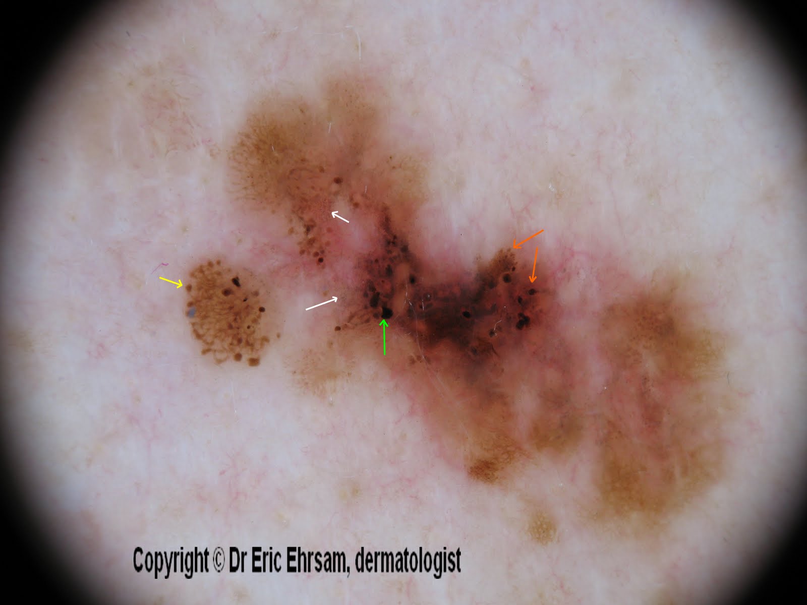
Back lesion


A 54-year-old woman consulted for this changing lesion on her back.


Dermoscopy revealed:

  • multiple colours, irregular pigmentation
  • a multi-component pattern
  • an atypical reticular pattern
  • irregular streaks (yellow arrow)
  • irregular dots and globules (orange arrow)
  • irregular blotches (green arrow)
  • peppering, regression (white arrow)


Pathology revealed a superficial spreading melanoma (SSM) with a Breslow index at 0.33 mm

Monday, 4 July 2011


A 25-year-old man consulted presented a pigmented lesion on his back (lumbar area).


Dermoscopy revealed:

  • a réticular pattern
  • irregular streaks (pseudopods)
in favor of a Spitz nevus which was confirmed after excision.

Saturday, 8 January 2011

Dermoscopy of lip pigmentation in a Laugier-Hunziker syndrome


Laugier-Hunziker syndrome is an acquired, benign, macular hyperpigmentation of buccal mucosa lips (and nails). Histologically, pigmentations are due to an accumulation of melanin in the basal layer keratinocytes and an increase in the number of melanophages in the submucosa and/or papillary dermis.

We report dermoscopic pictures of lip pigmentations in this syndrome.


In our case, dermoscopy revealed a reticular pattern and also linear or curvilinear streaks making a parallel pattern.


Reference:
Dermoscopic findings in Laugier-Hunziker syndrome. Gencoglan et al. Arch Dermatol. 2007; 143(5): 631-633


Monday, 16 March 2009

scalp melanoma


A 65-year-old man consulted for this pigmented lesion on his scalp.

Dermoscopy revealed a disorganised atypical reticular network, blue gray dots and regression, irregular branched streaks.
This lesion was excised and pathology revealed a melanoma in situ.


Saturday, 10 May 2008

Atypical nevus


A 32-year-old woman consulted for a mole check-up.
Clinical examination revealed the greatest mole localized on her left scapular area.
At dermoscopy, this lesion featured a pink veil, irregular streaks, irregular globules, an atypical reticular pattern, linear irregular vessels.


Irregular globules.

Polymorphous vessels including irregular linear vessels.

Irregular streaks.

Two diagnoses were possible: superficial spreading melanoma or atypical nevus. The patient did not mention any changing in surface or borders.
This lesion was excised and pathology revealed an atypical nevus ( compound) with a slight irritation.

Monday, 3 December 2007

Spitz nevus


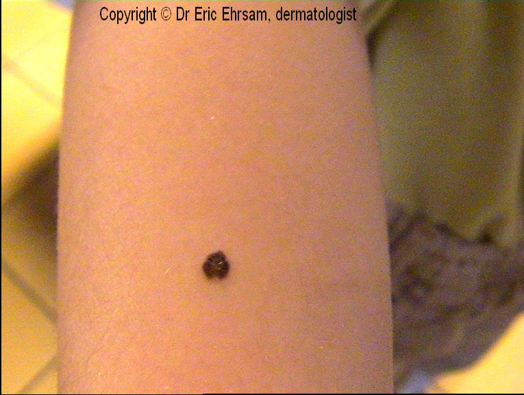
11-year-old boy with a dark pigmented lesion on his right arm.
Dermoscopy: symetric starburst pattern, streaks, regular globules at the center of the lesion. The diagnosis of Spitz nevus was made and a dermoscopic follow-up did not reveal enlargement or changing of structures.


Saturday, 1 December 2007

Superficial spreading melanoma


A 50-year-old man presented an irregularly pigmented lesion his back. Dermoscopy revealed an asymetric pigmentation, multiple colors, streaks (red circle), and a delicate blue white veil in favor of a superficial spreading melanoma.
The lesion was removed with a 2 mm margin and histology confirmed the diagnosis. The Breslow index was measured 0.63 mm. A re-excision was performed with a 1 cm margin. No mestastases were detected.


Other cases of melanoma
0 1 2 3 4 5 6 7 8 9 10 11 12 13 14 15 16 17 18