Search This Blog

Showing posts with label irregular globules. Show all posts

Friday, 12 February 2016

Brachial melanoma


A 54-year-old man had a rapidly growing lesion on his right back for 6 months.


Dermoscopy revealed:
  • a vascular pattern with some polymorphous and atypical vessels
  • peripheral pigmentation
  • irregular globules
  • eccentric blotches


Pathology revealed a superficial spreading melanoma with Breslow index at 1.53 mm

Saturday, 28 November 2015

Pigmented lesion on neck.


A 57 year-old-woman consulted for a pigmented lesion on her neck.


Dermoscopy revealed:
  • no melanocytic structures
  • a structureless pattern
  • irregular blue gray dots/globules 


Pathology was in favor of a Bowen's disease.

Friday, 27 November 2015

Thoracic pigmented lesion


Man 52 years old; Acquired pigmented lesion on chest.


Dermoscopy revealed:

  • multiple colors
  • asymmetry of form and structures
  • an atypical network
  • a blue white veil
  • irregular dots and globules
Pathology revealed a melanoma in situ.

Friday, 22 July 2011

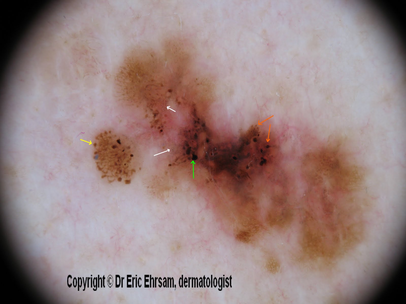
Back lesion


A 54-year-old woman consulted for this changing lesion on her back.


Dermoscopy revealed:

  • multiple colours, irregular pigmentation
  • a multi-component pattern
  • an atypical reticular pattern
  • irregular streaks (yellow arrow)
  • irregular dots and globules (orange arrow)
  • irregular blotches (green arrow)
  • peppering, regression (white arrow)


Pathology revealed a superficial spreading melanoma (SSM) with a Breslow index at 0.33 mm

Saturday, 7 May 2011

Forearm lesion


A 59-year-old woman consulted for this pigmented lesion on her right forearm.


Dermoscopy revealed:
  • irregular pigmentation
  • atypical reticular pattern
  • irregular dots / globules
  • brown dots
  • regressive homogeneous area
in favor of a melanoma.


This lesion was excised and pathology revealed a superficial spreading melanoma with a Breslow index at 0,47mm

Friday, 20 March 2009

Pigmented lesion on upper lip.

A 32 year-old woman consulted for this pigmented lesion present for 6 months.

Dermoscopy revealed:
  • an atypical pseudonetwork
  • rhomboidal structures
  • irregular globules
A melanoma was suspected and this lesion was excised. Pathology revealed a superficial spreading melanoma in situ (Clark level I).

Saturday, 10 May 2008

Atypical nevus


A 32-year-old woman consulted for a mole check-up.
Clinical examination revealed the greatest mole localized on her left scapular area.
At dermoscopy, this lesion featured a pink veil, irregular streaks, irregular globules, an atypical reticular pattern, linear irregular vessels.


Irregular globules.

Polymorphous vessels including irregular linear vessels.

Irregular streaks.

Two diagnoses were possible: superficial spreading melanoma or atypical nevus. The patient did not mention any changing in surface or borders.
This lesion was excised and pathology revealed an atypical nevus ( compound) with a slight irritation.

Monday, 17 March 2008

Early superficial spreading melanoma

A 37-year-old man consulted for an enlarging pigmented lesion on his abdomen.


Dermoscopy revealed an atypical pattern with an asymmetry, homogeneous area, irregular dots and globules, and irregular branched streaks.


The lesion was excised and pathology revealed a superficial spreading melanoma with a Breslow index non measurable.

Other cases of melanoma
0 1 2 3 4 5 6 7 8 9 10 11 12 13 14 15 16 17 18

Tuesday, 6 November 2007

Pseudopods

A 60-year-old man presented a nodular pigmented tumor on his chest. Dermoscopy revealed a pigment network at the periphery with many pseudopods (white circle), irregular dots and globules at 6 o'clock and 3 o'clock, and a blue white veil. The lesion was excised and pathology revealed superficial spreading melanoma with a Breslow index at 1.5mm and a Clark level II.

Other cases of melanoma
0 1 2 3 4 5 6 7 8 9 10 11 12 13 14 15 16 17 18

Monday, 13 August 2007

superficial spreading melanoma


This case concerned a 59-year-old patient: this pigmented lesion of his right thigh was discovered during a consultation for condylomas. The patient was not conscious of this lesion.

On dermoscopy (x 20) : Multicomponent pattern, irregular pigment network, blue white veil very irregular, irregular streaks (bottom) irregular dots and globules.


Other cases of melanoma
0 1 2 3 4 5 6 7 8 9 10 11 12 13 14 15 16 17 18